
Fig. 1
- ID
- ZDB-IMAGE-070919-4
- Publication
- Rau et al., 2006 - Zebrafish Trap230/Med12 is required as a coactivator for Sox9-dependent neural crest, cartilage and ear development
- All Figures
- Figures for Rau et al., 2006
|
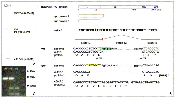
Fig. 1 Molecular characterization of the tpd locus. (A) tpd maps to linkage group 14 (LG 14), between markers Z53264 and Z11725, close to marker P1. (B) The tpd mutation (marked in red) causes an alteration of the splice donor site of intron 12. Two defectively spliced transcripts are generated as a result of this mutation. cDNA1 is spliced at a cryptic splice site (shaded in yellow) within exon 12. This leads to a frame shift and a stop codon after 93 amino acids. cDNA2 contains unspliced intron 12, leading to a stop codon after 4 amino acids. (C) Restriction enzyme Est1 cuts genomic tpd but not WT DNA due to the point mutation in its recognition site. het: DNA from a heterozygous embryo. L: leucine-rich domain; LS leucine-and-serine-rich domain; PQL: proline-, glutamine-, and leucine-rich domain; OPA: glutamine-rich domain.
Reprinted from Developmental Biology, 296(1), Rau, M.J., Fischer, S., and Neumann, C.J., Zebrafish Trap230/Med12 is required as a coactivator for Sox9-dependent neural crest, cartilage and ear development, 83-93, Copyright (2006) with permission from Elsevier. Full text @ Dev. Biol.