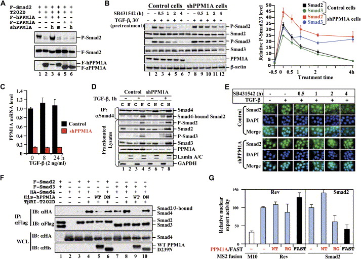
Fig. 4 PPM1A Regulates Phosphorylation, Oligomerization, and Nuclear Export of Smads (A) Specific PPM1A knockdown in 293T cells. Flag-hPPM1A, Flag-tagged human PPM1A; Flag-zPPM1A, Flag-zebrafish PPM1A; h, shPPM1A494 against human PPM1A; m, shmPPM1A494 against mouse PPM1A. The level of P-Smad2 was inversely correlated with that of PPM1A. (B) Reduced Smad2/3 dephosphorylation in HaCaT-shPPM1A cells. Left: Cells were treated with TGFβ and SB431542 and analyzed as described in Figure 1C. Right: Line graph showing relative levels of P-Smad2/3 over those of total Smad2/3 from three independent clones with two experiments each, with values and error bars representing the average and standard deviation. (C) qRT-PCR analysis of PPM1A mRNA in shPPM1A cells. Values and error bars represent mean and standard deviation of two experiments (each with triplicates). (D) Stable PPM1A depletion increases Smad2 phosphorylation and association with Smad4. HaCaT-shPPM1A or control cells were treated with TGFβ (2 ng/ml, 1 hr). Nuclear and cytoplasmic fractions (Supplemental Data) were subjected to anti-Smad4 IP and Western blotting. (E) PPM1A depletion increases Smad2 accumulation in the nucleus. Cells were treated with TGFβ for 1 hr and then with SB431542 for 0.5, 1, 2, or 4 hr. Smad2 was visualized using anti-Smad2 antibody (FITC). DAPI (DNA staining) and merge are indicated. (F) Wild-type PPM1A (WT), but not D239 mutant (DN), causes Smad2/3-Smad4 dissociation. (G) PPM1A promotes nuclear export of Smad2. An MS2-based quantitative analysis of nuclear transport assay system was used (Supplemental Data). Values and error bars represent mean and standard deviation of three experiments.
Reprinted from Cell, 125(5), Lin, X., Duan, X., Liang, Y.Y., Su, Y., Wrighton, K.H., Long, J., Hu, M., Davis, C.M., Wang, J., Brunicardi, F.C., Shi, Y., Chen, Y.G., Meng, A., and Feng, X.H., PPM1A functions as a Smad phosphatase to terminate TGFbeta signaling, 915-928, Copyright (2006) with permission from Elsevier. Full text @ Cell

