
Fig. 1
- ID
- ZDB-IMAGE-070918-52
- Publication
- Walker et al., 2006 - Zebrafish furin mutants reveal intricacies in regulating Endothelin1 signaling in craniofacial patterning
- All Figures
- Figures for Walker et al., 2006
|
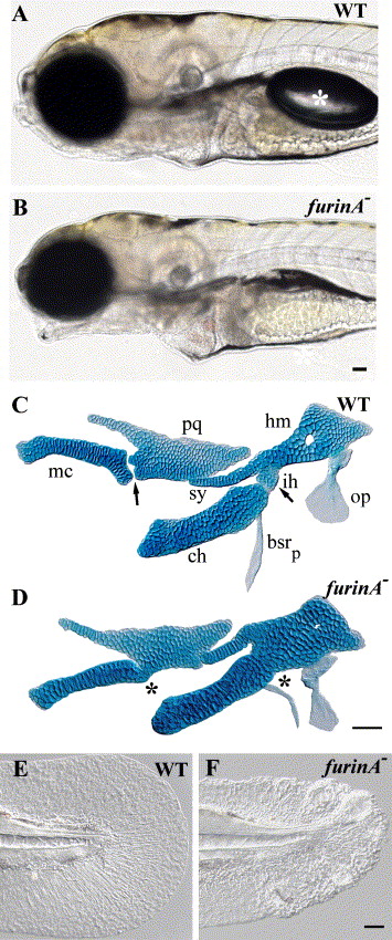
Fig. 1 furinA mutants have jaw and fin defects. (A and B) Live views at 5 dpf. furinA mutants have an open-mouth phenotype and fail to form a swim bladder (white asterisk in panel A). (C and D) Flat mounts of Alcian green-stained cartilages at 5 dpf. Wild-type DV joint regions are indicated with arrows in panel C. Fusions at joint regions in furinA mutants are indicated with asterisks in panel D. Cartilages are labeled as followed: pq (palatoquadrate), mc (Meckel's cartilage), hm (hyomandibula), ch (ceratohyal), sy (symplectic), and ih (interhyal). Two bones of the hyoid arch are also lightly stained with Alcian green: op (opercle), bsrp (branchiostegal ray posterior). (E and F) Live views of the tail fin at 2 dpf. furinA mutant larvae have mildly ruffled fins. Scale bars: 50 μm.
Reprinted from Developmental Biology, 295(1), Walker, M.B., Miller, C.T., Talbot, J.C., Stock, D.W., and Kimmel, C.B., Zebrafish furin mutants reveal intricacies in regulating Endothelin1 signaling in craniofacial patterning, 194-205, Copyright (2006) with permission from Elsevier. Full text @ Dev. Biol.