Image
Figure Caption
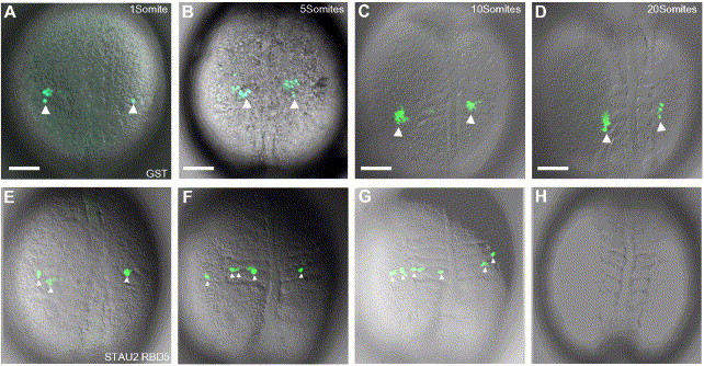
Fig. 7 PGC migration is aberrant when Stau2 function is disrupted. Time-lapse microscopy on GST-injected embryos (A–D) shows clusters of GFP:nos expressing cells (large white arrowheads) that migrate to the gonadal ridge. In contrast, in embryos injected with Stau2 RBD5 (E–H), GFP expressing cells fail to form clusters, remain dispersed (small white arrowheads in panels E, F, G), do not migrate to the gonadal ridge, and eventually disappear (H). Dorsal views of 1 somite stage (A, E), 5 somite stage (B, F), 10 somite stage (C, G), and 20 somite stage (D, H) embryos. Scale bars, 100 μm.
Acknowledgments
This image is the copyrighted work of the attributed author or publisher, and
ZFIN has permission only to display this image to its users.
Additional permissions should be obtained from the applicable author or publisher of the image.
Reprinted from Developmental Biology, 292(2), Ramasamy, S., Wang, H., Quach, H.N., and Sampath, K., Zebrafish Staufen1 and Staufen2 are required for the survival and migration of primordial germ cells, 393-406, Copyright (2006) with permission from Elsevier. Full text @ Dev. Biol.

