
Image
|
Figure Caption
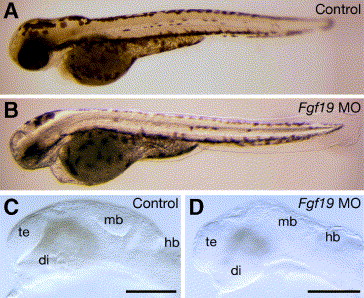
Fig. 3 Morphology of the Fgf19 MO-injected embryos. Embryos were injected with control Fgf19 MO (A and C) and Fgf19 MO (B and D). (A and B) 60 hpf. The Fgf19 MO-injected embryos had defects in the brain and eye. (C and D) 24 hpf. In the Fgf19 MO-injected embryos, the forebrain, midbrain and cerebellum were reduced in size. Lateral views with anterior to the left and dorsal to the top. di, diencephalon; hb, hindbrain; mb, midbrain; te, telencephalon. Scale bars, 30 μm.
Acknowledgments
This image is the copyrighted work of the attributed author or publisher, and
ZFIN has permission only to display this image to its users.
Additional permissions should be obtained from the applicable author or publisher of the image.
Reprinted from Developmental Biology, 288(1), Miyake, A., Nakayama, Y., Konishi, M., and Itoh, N., Fgf19 regulated by Hh signaling is required for zebrafish forebrain development, 259-275, Copyright (2005) with permission from Elsevier. Full text @ Dev. Biol.