
|
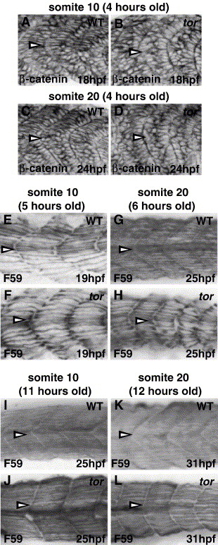
Fig. S2 Muscle fiber defects (elongation and slow fiber morphology) in tor mutant embryos are similar in anterior and posterior somites. Embryos were stained with β-catenin to compare fast muscle cell elongation in anterior (somite 10; A, B) and posterior (somite 20; C, D) somites. In wild type embryos, many fast muscle cells have initiated elongation 4 h after somite formation, regardless of position along the anterior–posterior axis (A, C; medial sections are 4–5 μM lateral to the notochord). In contrast, most tor mutant fast muscle cells at the same developmental stage remain mesenchymal, and the elongation delay is similar in somite 10 (anterior) and somite 20 (posterior) (B, D). (E–L) Slow muscle myosin (F59) expression reveals that slow muscle cell development is similar in anterior and posterior somites of tor mutant embryos. Approximately 5–6 h after somite formation, wild type slow muscle fibers reveal the basic metameric pattern (E, G), gaps appear in tor mutant somites, and the organization and morphology of slow muscle fibers appear similar in somite 10 and somite 20 (compare Panel F to H). Slow muscle defects recover in tor mutant somite/myotomes that formed 11–12 h earlier, and it is difficult to distinguish between wild type and mutant myotome based on slow muscle cell morphology alone (I–L), although tor mutant somite shape is distinctly U-shaped instead of chevron-shaped. All panels are lateral views with anterior to the left. Antibody and stage are indicated on each panel; somite number (indicated by arrowheads) and the age of that somite are indicated above each column.
Reprinted from Developmental Biology, 287(2), Dill, K.K., and Amacher, S.L., tortuga refines Notch pathway gene expression in the zebrafish presomitic mesoderm at the post-transcriptional level, 225-236, Copyright (2005) with permission from Elsevier. Full text @ Dev. Biol.