
Image
|
Figure Caption
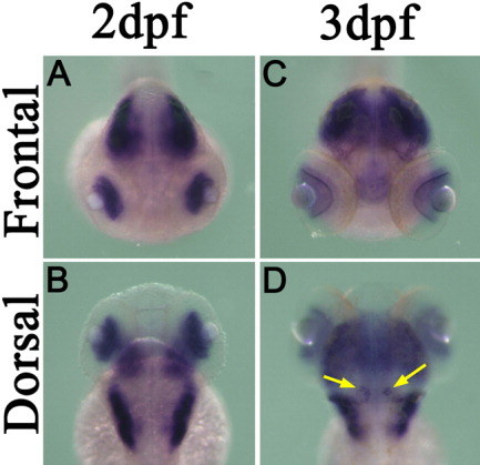
Fig. 4 Expression pattern of rora2 in the larval brain and eyes detected by whole-mount in situ hybridization. A-D: Frontal (A,C) and dorsal (B,D) views of 2 days postfertilization (dpf; A,B) and 3 dpf (C,D) specimens are shown. Frontal and dorsal views of a 3 dpf larva clearly show rora2 expression in the eyes and differentiating Purkinje cells (indicated by arrows in D), respectively.
Figure Data
Acknowledgments
This image is the copyrighted work of the attributed author or publisher, and
ZFIN has permission only to display this image to its users.
Additional permissions should be obtained from the applicable author or publisher of the image.
Full text @ Dev. Dyn.