
Image
|
Figure Caption
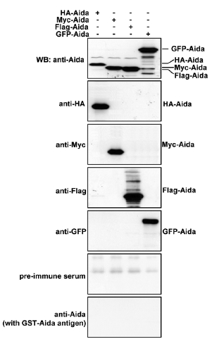
Fig. S2 Characterization of Aida polyclonal antibody. Differently tagged Aida plasmids were separately transfected into 293T cells. Protein samples from the differently transfected cells were subjected to immunoblotting with anti-HA, anti-Myc, anti-Flag, anti-GFP or anti-Aida antibodies to detect Aida expression. Anti-Aida recognized the same protein band as those detected by the respective tag antibodies. No specific band was detected with pre-immune serum or after blocking with Aida protein.
Acknowledgments
This image is the copyrighted work of the attributed author or publisher, and
ZFIN has permission only to display this image to its users.
Additional permissions should be obtained from the applicable author or publisher of the image.
Reprinted from Developmental Cell, 13(2), Rui, Y., Xu, Z., Xiong, B., Cao, Y., Lin, S., Zhang, M., Chan, S.C., Luo, W., Han, Y., Lu, Z., Ye, Z., Zhou, H.M., Han, J., Meng, A., and Lin, S.C., A beta-Catenin-Independent Dorsalization Pathway Activated by Axin/JNK Signaling and Antagonized by Aida, 268-282, Copyright (2007) with permission from Elsevier. Full text @ Dev. Cell