
Fig. 2
- ID
- ZDB-IMAGE-070822-18
- Publication
- Wakahara et al., 2007 - fibin, a novel secreted lateral plate mesoderm signal, is essential for pectoral fin bud initiation in zebrafish
- All Figures
- Figures for Wakahara et al., 2007
|
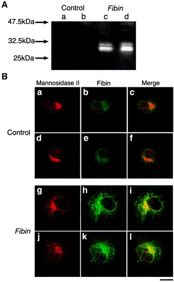
Fig. 2 Detection of recombinant mouse Fibin expressed in COS-7 cells. (A) COS-7 cells were transfected with the empty vector (Control) or the mouse Fibin-expression vector (Fibin). The cell lysate (a, c) and culture medium (b, d) of the transfected COS-7 cells were separated by SDS–polyacrylamide gel electrophoresis under reducing conditions and transferred onto a nitrocellulose membrane. The protein with the E tag on the membrane was detected using anti-E tag antibody. (B) COS-7 cells transfected with the empty vector (Control) (a–f) or the mouse Fibin-expression vector (Fibin) (g–l) were examined by immunohistochemistry using anti-mannosidase II antibody for the Golgi apparatus (a, d, g, j) and anti-E tag antibody for recombinant Fibin (b, e, h, k). The signals obtained by immunohistochemistry using anti-mannosidase II antibody and anti-E tag antibody were merged (c, f, i, l). Scale bar = 20 μm.
Reprinted from Developmental Biology, 303(2), Wakahara, T., Kusu, N., Yamauchi, H., Kimura, I., Konishi, M., Miyake, A., and Itoh, N., fibin, a novel secreted lateral plate mesoderm signal, is essential for pectoral fin bud initiation in zebrafish, 527-535, Copyright (2007) with permission from Elsevier. Full text @ Dev. Biol.