
IMAGE
Fig. S1
- ID
- ZDB-IMAGE-070821-97
- Publication
- Hall et al., 2006 - An essential role for zebrafish Fgfrl1 during gill cartilage development
- All Figures
- Figures for Hall et al., 2006
Image
|
Figure Caption
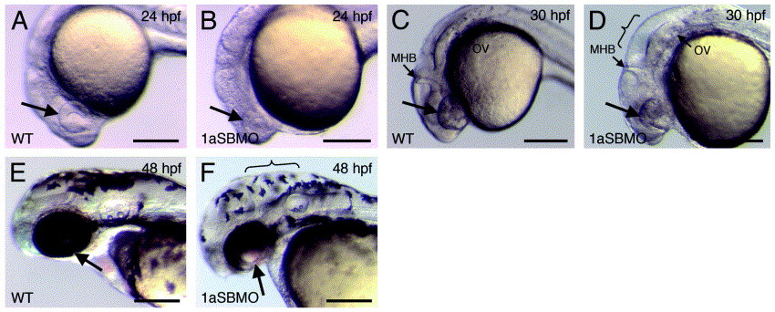
Fig. S1 Depletion of Fgfrl1a results in specific developmental defects. (A, C, and E) 24, 30 and 48 hpf wildtype embryos, respectively. (B, D and F) 24, 30 and 48 hpf Fgfrl1a morphant embryos, respectively (lateral views). Arrows mark developing eye. Braces mark expanded hindbrain region. Abbreviations: MHB, midbrain–hindbrain boundary; OV, otic vesicle. Scale bars: 250 μm.
Figure Data
Acknowledgments
This image is the copyrighted work of the attributed author or publisher, and
ZFIN has permission only to display this image to its users.
Additional permissions should be obtained from the applicable author or publisher of the image.
Reprinted from Mechanisms of Development, 123(12), Hall, C., Flores, M.V., Murison, G., Crosier, K., and Crosier, P., An essential role for zebrafish Fgfrl1 during gill cartilage development, 925-940, Copyright (2006) with permission from Elsevier. Full text @ Mech. Dev.