
|
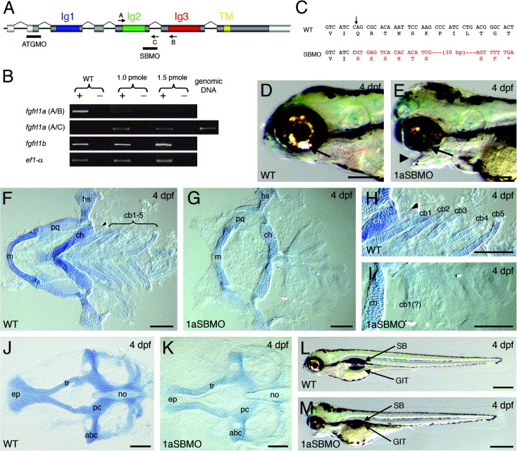
Fig. 4 Targeted depletion of Fgfrl1a results in specific defects within the pharyngeal cartilage. (A) Genomic organization of fgfrl1a illustrating the Ig-like (Ig1, 2 and 3) and transmembrane (TM) domains, and the target site for the splice-blocking (SBMO) and ATG (ATGMO) morpholinos. ORF and untranslated regions highlighted by dark and light shading, respectively. The location and orientation of primers used for RT-PCR detection of altered splicing are indicated by arrows. (B) Semi-quantitative RT-PCR illustrating specific depletion of normal fgfrl1a splicing following early delivery of 1.0 and 1.5 pmol doses of SBMO (+, reverse transcriptase present; -, reverse transcriptase absent). Primer combinations are bracketed. (C) Predicted translation of fgfrl1a following SBMO-mediated defective splicing. Arrow denotes splice site between Ig2- and Ig3-encoding exons. Black and red text corresponds to coding and intronic sequence, respectively. (D, F, H, J and L) 4 dpf wildtype embryos. (E, G, I, K and M) 4 dpf Fgfrl1a morphant embryos. (D/E) lateral views. (F–K) Dissected and flat-mounted cartilaginous elements stained with alcian blue. (F/H and G/I) Ventral cartilage elements from wildtype and Fgfrl1a morphant larvae, respectively. (J and K) Neurocranium from wildtype and Fgfrl1a morphant larvae, respectively. Arrowhead marks dysmorphic jaw. Abbreviations: abc, anterior basicranial commissure; cb1–5, ceratobranchial arch 1–5; cb1(?), presumptive remnants of ceratobranchial element 1; ch, ceratohyal; ep, ethmoid plate; GIT, gastrointestinal tract; hs, hyosymplectic; m, Meckel’s cartilage; no, notochord; pc, parachordal; pq, palatoquadrate; SB, swim bladder; tr, trabecula. Scale bars: 250 μm in D and E; 100 μm in F–K; 500 μm in L and M.
Reprinted from Mechanisms of Development, 123(12), Hall, C., Flores, M.V., Murison, G., Crosier, K., and Crosier, P., An essential role for zebrafish Fgfrl1 during gill cartilage development, 925-940, Copyright (2006) with permission from Elsevier. Full text @ Mech. Dev.