
Fig. 3
- ID
- ZDB-IMAGE-070821-91
- Genes
- Publication
- Hall et al., 2006 - An essential role for zebrafish Fgfrl1 during gill cartilage development
- All Figures
- Figures for Hall et al., 2006
|
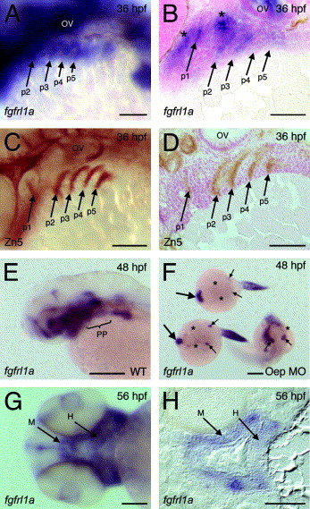
Fig. 3 Pharyngeal arch expression of fgfrl1a. (A and B) Lateral view of fgfrl1a expression within pharyngeal region of 36 hpf embryo and equivalent sagittal section, respectively. Asterisks mark expression within arch 1- and 2-associated neural crest. (C and D) Lateral view of pharyngeal region of Zn5-stained embryo and equivalent sagittal section, respectively. (E) Lateral view of fgfrl1a expression within head of 48 hpf embryo. (F) Expression of fgfrl1a in 48 hpf embryos following morpholino-mediated depletion of Oep (two embryos to left phenocopy maternal-zygotic Oep mutants while the embryo on the right a less severe, zygotic, Oep mutant). Large and small arrows denote persistence of fgfrl1a transcript in anterior domains and otic vesicles, respectively. Asterisks highlight absence of expression in pharyngeal pouch endoderm. (G and H) Ventral view of head region at 56 hpf and equivalent frontal section, respectively. Abbreviations: H, hyoid arch; M, mandibular arch; OV, otic vesicle; p1–5, pharyngeal pouch 1–5; PP, pharyngeal pouches. Scale bars: 100 μm in A–D, G and H; 250 μm in E and F.
Reprinted from Mechanisms of Development, 123(12), Hall, C., Flores, M.V., Murison, G., Crosier, K., and Crosier, P., An essential role for zebrafish Fgfrl1 during gill cartilage development, 925-940, Copyright (2006) with permission from Elsevier. Full text @ Mech. Dev.