
|
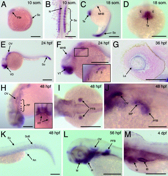
Fig. 2 Expression of fgfrl1a during zebrafish development. (A) Lateral view of 10 somite stage embryo. (B) Flat mount of 10 somite stage embryo. (C) Lateral view of 18 somite stage embryo. (D) Higher magnification of forebrain region at 18 somite stage. (E) Lateral view of 24 hpf embryo. (F) Higher magnification of cranio-trunk region at 24 hpf; inset represents enlarged view of boxed region. Pharyngeal pouches marked as 1, 2 and 3. (G) Section through eye at 36 hpf. (H) Dorsolateral view of head region of 48 hpf embryo; asterisks mark pharyngeal pouches, inset represents frontal view of head region. (I) Dorsal view of anterior trunk region of 48 hpf embryo. (J) Dorsolateral view of anterior trunk at 48 hpf. (K) Lateral view of tail region of 48 hpf embryo. (L) Lateral view of head region at 56 hpf. (M) Lateral view of anterior trunk at 4 dpf. Abbreviations: An, anus; E, eye; Ec, ectoderm; FBr, forebrain; H, hyoid arch; HT, heart tube; IB, intestinal bulb; IBP, intestinal bulb primordium; Le, lens; Li, liver; M, mandibular arch; MHB, midbrain–hindbrain boundary; N, notochord; OV, otic vesicle; PFB, pectoral fin bud; PP, pharyngeal pouches; So, somites; SoB, somite boundaries; Te, telencephalon; VD, ventral diencephalon; VT, ventral telencephalon. Scale bars: 250 μm in A–F, H, I and K–M; 100 μm in G and J.
Reprinted from Mechanisms of Development, 123(12), Hall, C., Flores, M.V., Murison, G., Crosier, K., and Crosier, P., An essential role for zebrafish Fgfrl1 during gill cartilage development, 925-940, Copyright (2006) with permission from Elsevier. Full text @ Mech. Dev.