
Fig. 8
- ID
- ZDB-IMAGE-070821-17
- Publication
- Wallace et al., 2005 - Intestinal growth and differentiation in zebrafish
- All Figures
- Figures for Wallace et al., 2005
|
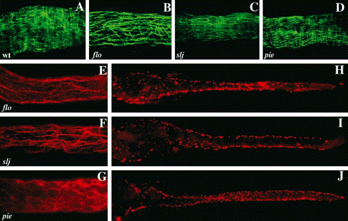
Fig. 8 Circular smooth muscle and enteric neuron defects accompany epithelial alterations in slj and flo, but not in pie mutants. (A–D) Whole-mount view of a segment of the mid intestine of WT, flo, slj and pie larvae processed for desmin immunohistochemistry. The WT intestine (A) contains short longitudinal smooth muscle fibers and longer, underlying circular smooth muscle fibers. In the flo (B) and slj (C) intestine the longitudinal fibers predominate. By contrast, smooth muscle appears normal in pie larvae (D). (E–G) Axonal projections of enteric neurons in the mid intestine of 5 dpf flo, slj and pie larvae. Axonal projections of flo (E) and slj (F) are less complex than WT (Fig. 6L) even at 96 hpf. Axonal projections are predominantly aligned along the anterior–posterior axis in both mutants. (G) Axonal projections in pie larvae appear normal. (H–J) Whole-mount images of 5 dpf flo, slj and pie larvae processed for Hu immunohistochemistry. Compared with WT larvae (Fig. 6I,K) fewer flo and slj enteric neurons have migrated from the lateral intestinal borders, whereas the pie pattern of Hu+ cells more closely resembles WT (Fig. 6K). The number of Hu+ cells is reduced in flo and slj and fewer flo Hu+ cells have migrated to the posterior intestinal segment.
Reprinted from Mechanisms of Development, 122(2), Wallace, K.N., Akhter, S., Smith, E.M., Lorent, K., and Pack, M., Intestinal growth and differentiation in zebrafish, 157-73, Copyright (2005) with permission from Elsevier. Full text @ Mech. Dev.