
Fig. 3
- ID
- ZDB-IMAGE-070821-10
- Publication
- Schorpp et al., 2002 - A zebrafish orthologue (whnb) of the mouse nude gene is expressed in the epithelial compartment of the embryonic thymic rudiment
- All Figures
- Figures for Schorpp et al., 2002
|
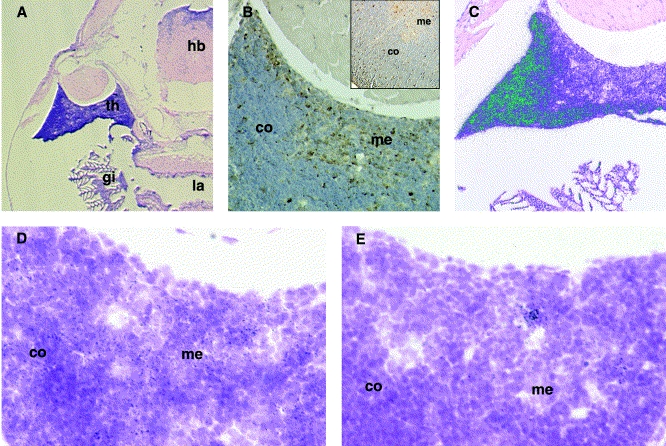
Fig. 3 Characterization of adult zebrafish thymus tissue. (A) Haematoxylin–eosin staining of a zebrafish thymus 6 weeks after fertilization. Note the darkly stained thymus (th) adjacent to the gill chamber (gi). la, larynx; hb, hindbrain. (B) Binding pattern of ConA lectin in zebrafish thymus; inset, adult mouse thymus. The presumptive cortical (co) and medullary (me) regions are indicated. (C) RNA in situ hybridization with radio-labelled rag-1 probe; silver grains are displayed in false colour (green). (D) Analysis of whnb in thymus section at the cortico-medullary junction. Note the equal distribution of silver grains (black dots). (E) Analysis of whna in thymus section at the cortico-medullary junction. Note that expression is confined to a few cells.
Reprinted from Mechanisms of Development, 118(1-2), Schorpp, M., Leicht, M., Nold, E., Hammerschmidt, M., Haas-Assenbaum, A., Wiest, W., and Boehm, T., A zebrafish orthologue (whnb) of the mouse nude gene is expressed in the epithelial compartment of the embryonic thymic rudiment, 179-185, Copyright (2002) with permission from Elsevier. Full text @ Mech. Dev.