
|
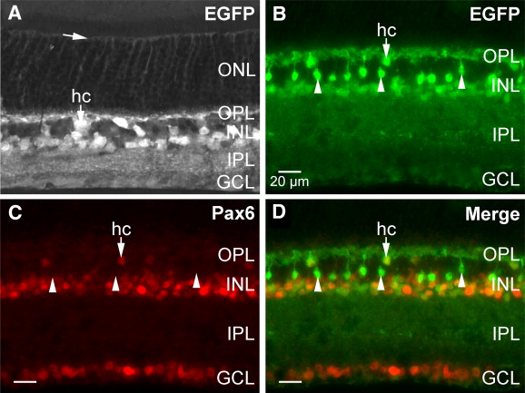
Fig. 8 Zebrafish Pax6a P0/P1 promoters are active in Pax6-expressing cells in the adult mouse retina. (A–B) EGFP from the zebrafish Pax6a BAC transgene is expressed by cells in the inner nuclear layer (INL) and ganglion cell layer (GCL). (A) EGFP+ radial processes also span the outer nuclear layer (ONL) and end at the outer limiting membrane (arrow). This suggests that Müller glia express the zebrafish Pax6a BAC transgene. (B–D) Direct comparison of EGFP and Pax6 expression in the adult mouse retina revealed that most, if not all, Pax6+ cells coexpressed EGFP; however, a few EGFP+ cells did not express Pax6 (arrowheads). The ectopic expression in these cells is most likely due to either species differences in expression or misregulation of the zebrafish Pax6a BAC transgene in mice. Scale bar in panel B applies to panels C–D.
Reprinted from Developmental Biology, 307(2), Lakowski, J., Majumder, A., and Lauderdale, J.D., Mechanisms controlling Pax6 isoform expression in the retina have been conserved between teleosts and mammals, 498-520, Copyright (2007) with permission from Elsevier. Full text @ Dev. Biol.