
Fig. 1
- ID
- ZDB-IMAGE-070802-28
- Publication
- Kok et al., 2007 - The role of the SPT6 chromatin remodeling factor in zebrafish embryogenesis
- All Figures
- Figures for Kok et al., 2007
|
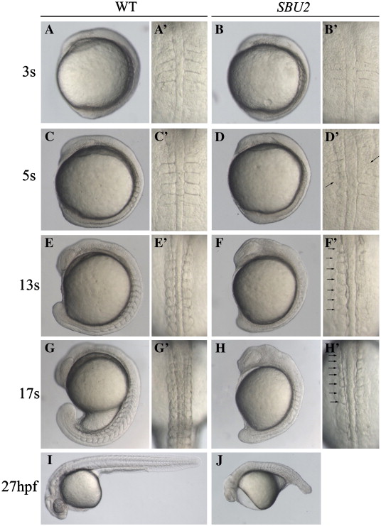
Fig. 1 Phenotype of SBU2 mutants. (A–J) Lateral views of living WT and SBU2 embryos, anterior to the top and dorsal to the right. (A′–H′) Dorsal views of panels A–H, anterior to the top. At early somitogenesis, somite boundaries are irregular and indefinite (A′ and B′) in SBU2 embryos. At ∼ 5-somite stage, somites are formed asymmetrically while previously formed somite boundaries start to degenerate (arrows) (C′ and D′). By the 5-somite stage SBU2 mutants have a slightly shorter body length then their wild-type siblings (C and D) and by the 13-somite stage, SBU2 mutants are clearly less extended along A–P axis than wild-type embryos (E and F). While somites continue to form in wild-type embryos, SBU2 mutants have only irregularly formed 6–7 somites and lack posterior somites (arrows) (F′ and H′). By 24–27 hpf, most of the previously formed structures including somite boundaries are degraded or distorted and the embryos degenerate (J). Note: all of the embryos shown in this figure and the following figures are diploids.
Reprinted from Developmental Biology, 307(2), Kok, F.O., Oster, E., Mentzer, L., Hsieh, J.C., Henry, C.A., and Sirotkin, H.I., The role of the SPT6 chromatin remodeling factor in zebrafish embryogenesis, 214-226, Copyright (2007) with permission from Elsevier. Full text @ Dev. Biol.