
Fig. 4
- ID
- ZDB-IMAGE-070619-20
- Publication
- Qian et al., 2007 - Distinct Functions for Different scl Isoforms in Zebrafish Primitive and Definitive Hematopoiesis
- All Figures
- Figures for Qian et al., 2007
|
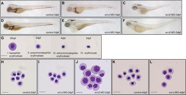
Fig. 4 Requirement of scl-α and -β at Different Development Stages of Primitive RBC Differentiation (A–F) O-dianisidine staining of hemoglobin. At 3 dpf, expression of hemoglobin was severely reduced in scl-β morphants (C) compared to control embryos (A) and scl-α morphants (B). At 5 dpf, the o-dianisidine staining in both scl-α (E) and scl-β (F) morphants was dramatically decreased compared to that in control embryos (D). All embryos are in lateral view with anterior to the left. (G–L) May-Grunwald Giemsa staining of RBCs of different developmental stages (30 hpf to 5 dpf). Normal primitive RBCs at 30 hpf to 5 dpf can be classified into four main developmental stages (G): stage I, basophilic erythroblast; stage II, polychromatophilic erythroblast; stage III, orthochromatophilic erythroblast; and stage IV, mature erythrocyte. While RBCs from 2-dpf control embryos (H) and scl-α morphants (I) have differentiated into the polychromatophilic erythroblast stage (stage II), RBCs from scl-β morphants (J) are halted at the earlier basophilic erythroblast stage (stage I). At 4 dpf, RBCs from scl-α morphants (L) are arrested at stage II, while there is normal development of stage IV RBCs in control embryos (K). Scale bar (G–L), 10 μm.