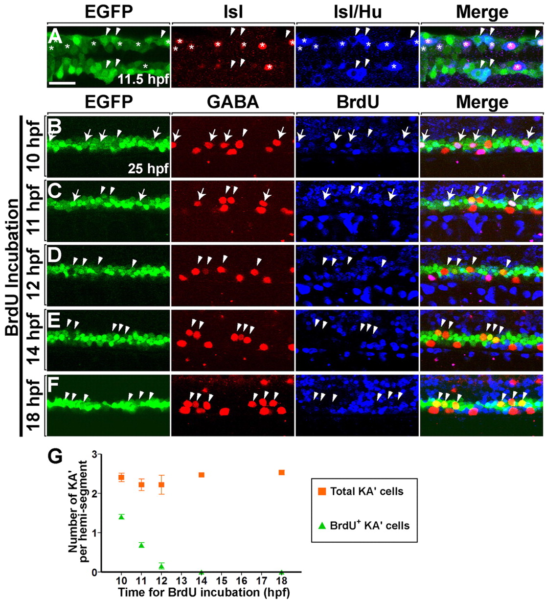
Fig. 1 Neural plate olig2:EGFP+ precursors generate PMNs and KA' interneurons at the same time. (A) Dorsal view of the posterior neural plate at 11.5 hpf (five-somite stage) and (B-F) lateral view of 6- to 12-somite regions of spinal cord at 25 hpf in Tg(olig2:egfp) zebrafish embryos. (A) Heterogeneous primary neuronal populations within the EGFP+ domain. Asterisks and arrowheads mark EGFP+ Hu+ Isl+ PMNs and EGFP+ Hu+ Isl- interneurons, respectively. Isl protein is nuclear, whereas Hu is cytoplasmic. (B-F) Embryos were incubated with BrdU at successive timepoints (as shown to the left of each panel) and labeled with anti-GABA (red) and anti-BrdU (blue) antibodies. Arrows and arrowheads mark BrdU+ and BrdU- KA' interneurons, respectively. (B) Four KA' interneurons were formed from EGFP+ precursors that underwent S phase at 10 hfp (arrows). Arrowhead marks BrdU- KA' interneuron, indicating that the postmitotic cell was formed before or after 10 hpf. (C) Two BrdU+ KA' interneurons (arrows) and two BrdU- KA' interneurons (arrowheads) were detected in the embryos that were incubated with BrdU at 11 hpf. (D,E,F) Embryos incubated with BrdU at 12, 14 and 18 hpf. No BrdU+ KA' interneurons were evident. (G) Average of all KA' cells (squares) versus BrdU+ KA' cells (triangles) (n=4 animals each). S phase for the last KA' interneurons produced occurs between 10 and 12 hpf. Error bars represent s.e.m. Scale bar: 25 µm.
Image
Figure Caption
Acknowledgments
This image is the copyrighted work of the attributed author or publisher, and
ZFIN has permission only to display this image to its users.
Additional permissions should be obtained from the applicable author or publisher of the image.
Full text @ Development

