
IMAGE
Fig. 2
- ID
- ZDB-IMAGE-070501-3
- Publication
- Baye et al., 2007 - The disarrayed mutation results in cell cycle and neurogenesis defects during retinal development in zebrafish
- All Figures
- Figures for Baye et al., 2007
Image
|
Figure Caption
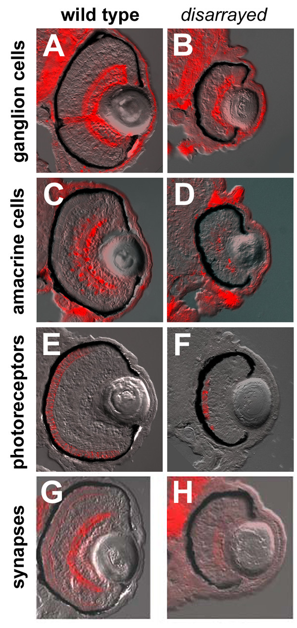
Fig. 2 Retinal cell-type marker analysis in disarrayed eyes. Transverse central retinal sections from 96 hpf wild-type (A,C,E,G) and disarrayed (B,D,F,H) embryos assessed for cell-type and lamina specific markers. (A,B) Immunofluorescence for zn8 antigen (retinal ganglion cells and their axons); (C, D) parvalbumin (amacrine cells and their processes); (E,F) zpr1 antigen (cone photoreceptor cells); and (G,H) SV2 antigen (synaptic vesicles). All cell types are present and in the correct laminar position. All figures are bright field images overlaid with the immunofluorescent label.
Figure Data
Acknowledgments
This image is the copyrighted work of the attributed author or publisher, and
ZFIN has permission only to display this image to its users.
Additional permissions should be obtained from the applicable author or publisher of the image.
Full text @ BMC Dev. Biol.