
IMAGE
Fig. 1
- ID
- ZDB-IMAGE-070501-2
- Publication
- Baye et al., 2007 - The disarrayed mutation results in cell cycle and neurogenesis defects during retinal development in zebrafish
- All Figures
- Figures for Baye et al., 2007
Image
|
Figure Caption
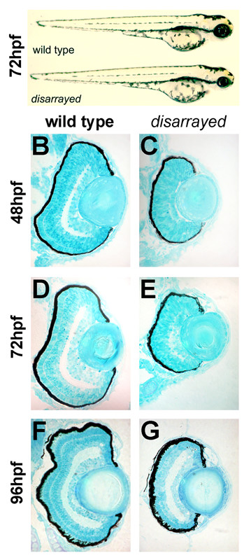
Fig. 1 Delayed retinal lamination in disarrayed eyes. (A) Lateral views of wild-type (upper) and disarrayed (lower) sibling embryos at 72 hpf. Mutant embryos are characterized by smaller eyes and forebrain by 42 hpf. Transverse central retinal sections of wild-type (B,D, F) and disarrayed (C,E,G) at 48 hpf (B,C), 72 hpf (D,E), and 96 hpf (F,G). Note the significant delay in differentiation and lamination in the disarrayed retina, although lens growth appears normal.
Figure Data
Acknowledgments
This image is the copyrighted work of the attributed author or publisher, and
ZFIN has permission only to display this image to its users.
Additional permissions should be obtained from the applicable author or publisher of the image.
Full text @ BMC Dev. Biol.