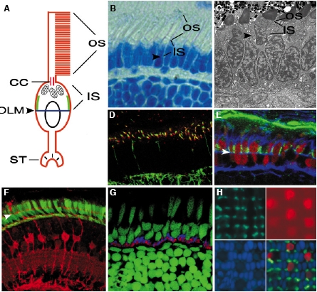
Fig. 1 The photoreceptor cell. (A) A schematic representation of the vertebrate photoreceptor cell. The photosensitive part of the photoreceptor cell, the outer segment, is a tall stack of membrane folds that connects to the rest of the photoreceptor body via a primary cilium, known as the connecting cilium. The basal end of the connecting cilium is anchored in the inner segment, the apical-most part of the photoreceptor perikaryal region, characterized by the presence of numerous mitochondria. The basal-most region of the photoreceptor cell features the synaptic terminus, which contains specialized synaptic junctions, called ribbon synapses. Similar to epithelial cells from which photoreceptors originate, the photoreceptor cell body is subdivided by a belt of cell junctions (OLM) into apical and baso-lateral domains. Distinct molecular properties of these membrane domains are evident in the distribution of several polypeptides, such as the nok or has gene products (green lines). (B) At 5 dpf, photoreceptor cells of the zebrafish larva feature long outer segments and are functional by electrophysiological criteria. In this panel, photoreceptor cells and their outer segments are visualized by methylene blue-staining of a plastic section. The retina shown in this panel was treated with PTU, a chemical that inhibits pigmentation revealing the presence of outer segments. (C) Inner segments are characterized by the presence of numerous mitochondria. They are evident on this TEM image of a section through the photoreceptor cell layer at 3 dpf. (D) Photoreceptor connecting cilia are visualized with antiacetylated tubulin antibodies (green). The IFT52 polypeptide, a component of the intraflagellar transport particle, is particularly highly concentrated at the base of cilia. Its presence is revealed by antibody staining (red). (E) Double cones of the zebrafish retina are visualized by antibody staining (red). Similar to other photoreceptor types, their surface is subdivided into apical and baso-lateral domains by a belt of adherens junctions, here visualized with fluorophore-conjugated phalloidin (blue). Antibody staining reveals that the Nagie oko polypeptide localizes next to adherens junctions in a narrow apical region of the photoreceptor surface (green). (F) The processes of Muller glia, here revealed by staining with anti-carbonic anhydrase antibody (red), extend into the photoreceptor cell layer and contribute to the junctions of the outer limiting membrane that subdivide the surface of photoreceptor cells (green). (G) Photoreceptor synapses are visualized with antibodies against syntaxin 3 (red) and SV2 (blue). (H) In the plane of the photoreceptor cell layer, cells form a regular pattern. Clockwise, starting from the lower left image: photoreceptor nuclei stained with DAPI (blue), rod opsin-GFP transgene expression (green), anti-UV-opsin antibody staining of short single cones (red), and a merged image of the previous three. CC, connecting cilium; IS, inner segment; OLM, outer limiting membrane; OS, outer segment, ST, synaptic terminus. In B through G, retinal pigment epithelium is up. Arrowheads indicate the approximate position of the outer limiting membrane. Panel H provided courtesy of Jim Fadool.
Image
Figure Caption
Acknowledgments
This image is the copyrighted work of the attributed author or publisher, and
ZFIN has permission only to display this image to its users.
Additional permissions should be obtained from the applicable author or publisher of the image.
Full text @ Int. J. Dev. Biol.

