
|
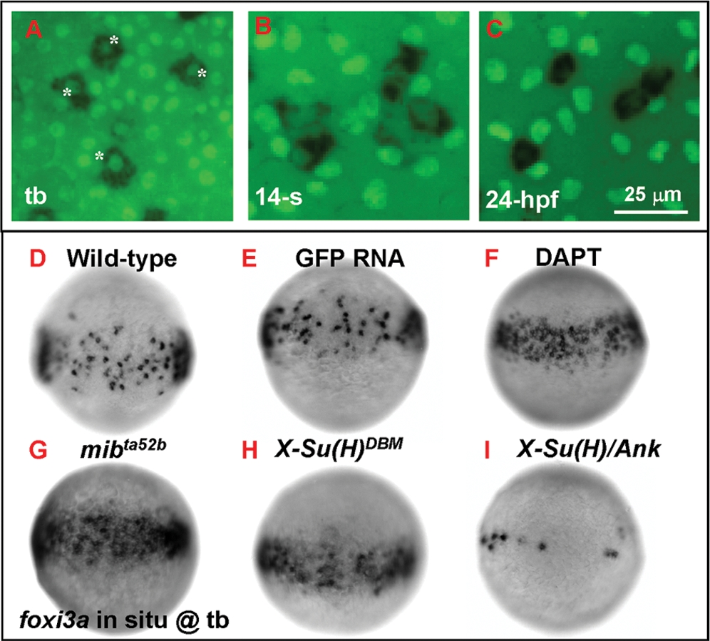
Fig. 2 Number of Epidermal Ionocyte Progenitors is Restricted by Delta-Notch-Mediated Lateral Inhibition. (A–C) Dynamic expression of the epidermal ionocyte progenitor marker of foxi3a (black, cytoplasmic staining) and the epidermal stem cell marker of P63 (green, nuclear staining) on the zebrafish epidermis. In the beginning, foxi3a was activated in some P63-positive epidermal stem cells at the tail bud (tb) stage (indicated by asterisks, A). However, as development precedes, P63 expression in epidermal ionocyte progenitors is sharply downregulated (at the 14-somite (14-s) stage, B) ultimately to an undetectable level (24 hour post infection (hpf), C). (D–I) The mosaic distribution of epidermal ionocyte progenitors within the epidermal ionocyte domain is controlled by Delta-Notch-mediated lateral inhibition. In DAPT-treated embryos (F), and mibta52b- (G) or X-Su(H)DBM mRNA-injected embryos (H), the foxi3a expression in the epidermal ionocyte domain has become denser and more homogeneous. While in X-Su(H)/Ank mRNA-injected embryos (I), foxi3a expression in the epidermal ionocyte domain is greatly reduced. The developmental stage is indicated in the left lower corner of each panel.