
|
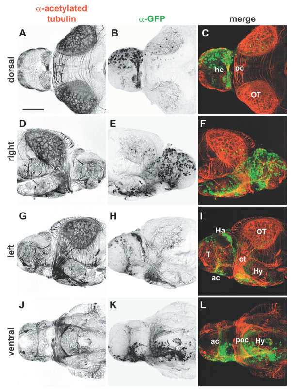
Fig. 4 Whole mount immunocytochemistry of electroporated brains. Isolated 4 dpf brains from embryos in which pHuC:GAL4/pUAS:EGFP were unilaterally electroporated into the right forebrain. Brains were stained with (a,d,g,j) anti-acetylated tubulin to label axons (red) and (b,e,h,k) anti-GFP to mark transfected cells (green); (c,f,i,l) merged images. Widespread expression of EGFP is seen in neuronal cell bodies of the right telencephalon and diencephalon (b,e). All major commissures contain labeled fibers and contralateral axonal projections can be seen in detail in left and ventral views (h,k). Anterior is to right in (d-f), to the left in all other panels. Scale bar = 100 µm. Ac, anterior commissure; Ha, habenula; hc, habenular commissure; Hy, hypothalamus; ot, optic tract; OT, optic tectum; pc, posterior commissure; poc, postoptic commissure; T, telencephalon.