
Fig. S6
- ID
- ZDB-IMAGE-070413-15
- Publication
- Hsiao et al., 2007 - A Positive Regulatory Loop between foxi3a and foxi3b Is Essential for Specification and Differentiation of Zebrafish Epidermal Ionocytes
- All Figures
- Figures for Hsiao et al., 2007
|
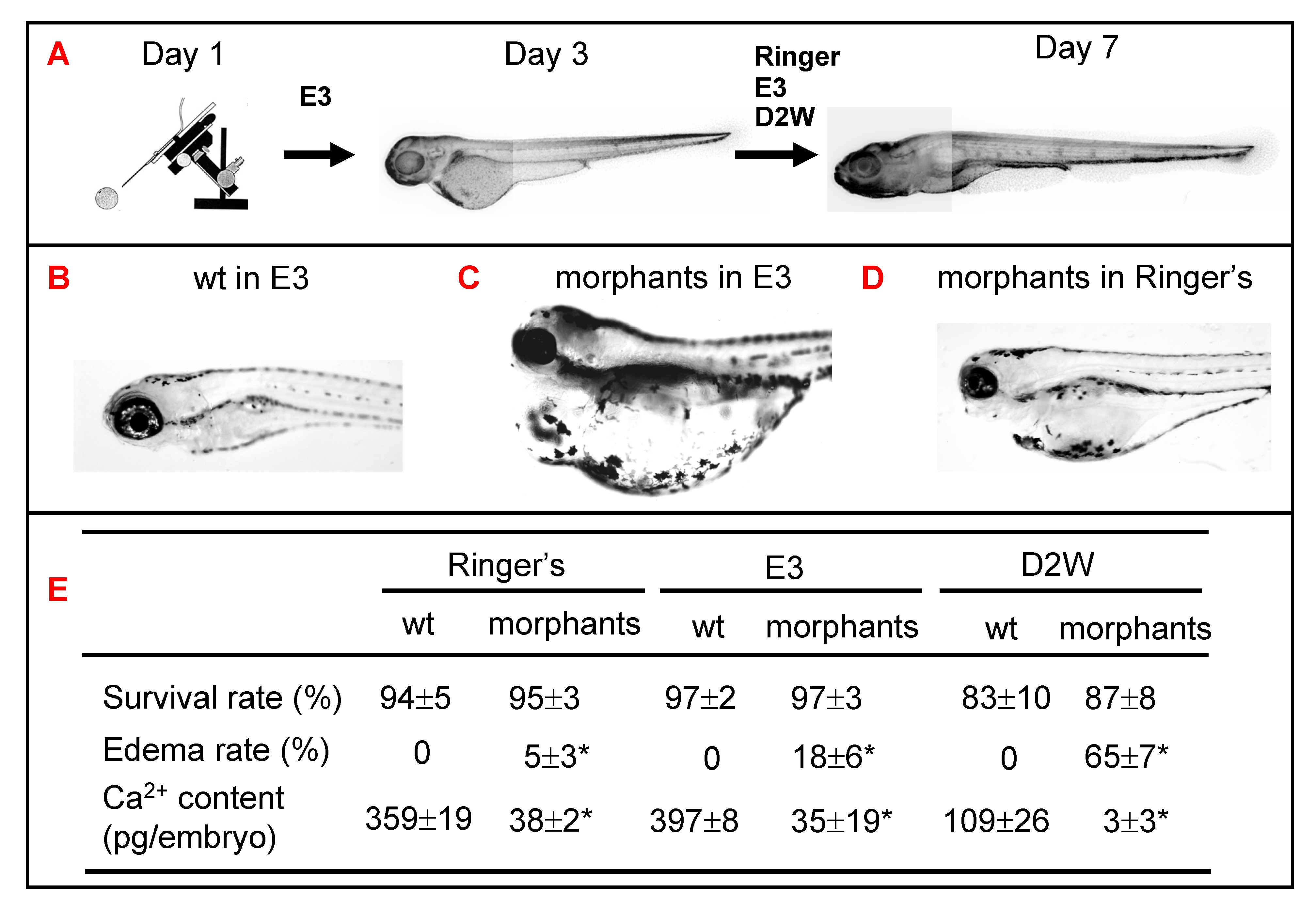
Fig. S6 Role of epidermal ionocytes in water and ion homeostasis in zebrafish embryos. (A) Procedure to assay the physiological functions of epidermal ionocytes in zebrafish embryos. After injecting them with foxi3a MO, morphants were initially raised in E3 up to 3 days post-fertilization (dpf) and then challenged with either Ringer's solution, E3, or double-distilled water (D2W). The survival rate, edema rate, and whole-body Ca2+ content between wild-types (wt) and morphants were measured at 7 dpf, and results are summarized in (E). The wild-types had a strong water balance ability and showed no edema phenotype in either E3 (B), Ringer's solution, or D2W (not shown). The morphants displayed a severely edematous phenotype in hypotonic E3 (C) or D2W (not shown), while the abnormality was greatly rescued in isotonic Ringer's solution (D). The values are shown as the mean±SD (n = 10). Asterisks (*) indicate a significant difference from the wild-type (Student's t-test, p<0.05).