
IMAGE
Fig. S1
- ID
- ZDB-IMAGE-070413-10
- Publication
- Hsiao et al., 2007 - A Positive Regulatory Loop between foxi3a and foxi3b Is Essential for Specification and Differentiation of Zebrafish Epidermal Ionocytes
- All Figures
- Figures for Hsiao et al., 2007
Image
|
Figure Caption
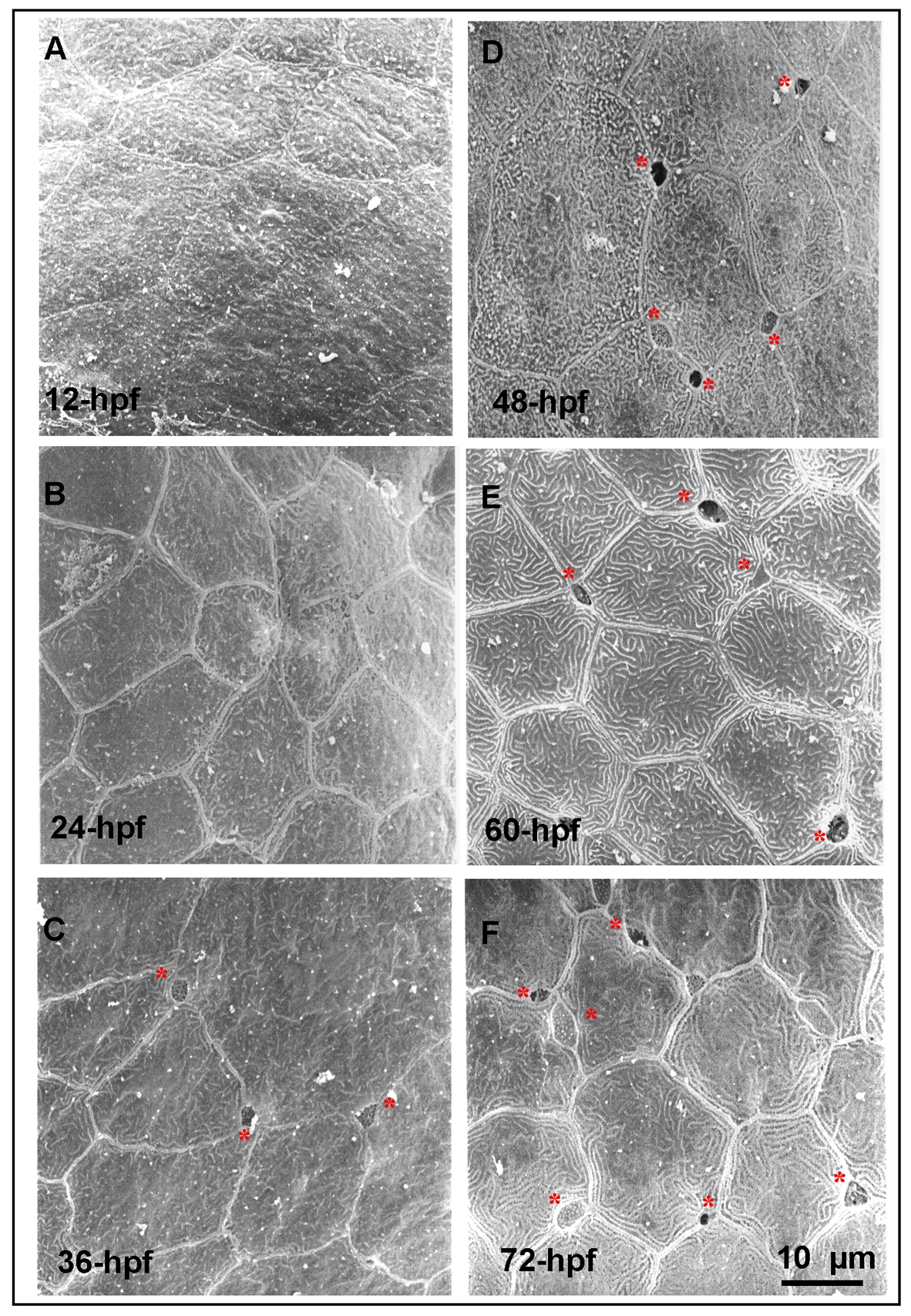
Fig. S1 Detection of the apical opening of epidermal ionocytes in zebrafish embryos.(A-F) The epidermal layer covering the yolk ball of wild-type embryos was scanned by a scanning electron microscope at different developmental stages (indicated in the lower left-hand corner). The first apical opening of the epidermal ionocyte appeared at 36 hours post-fertilization (hpf) (asterisks).
Acknowledgments
This image is the copyrighted work of the attributed author or publisher, and
ZFIN has permission only to display this image to its users.
Additional permissions should be obtained from the applicable author or publisher of the image.
Full text @ PLoS One