
Fig. 4
- ID
- ZDB-IMAGE-070309-8
- Publication
- Wilson et al., 2007 - Cadherin-4 plays a role in the development of zebrafish cranial ganglia and lateral line system
- All Figures
- Figures for Wilson et al., 2007
|
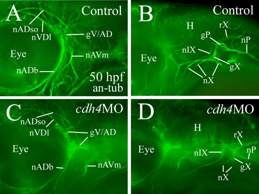
Fig. 4 Cranial and lateral line ganglion nerves in a control embryo (A,B) and an embryo injected with RcadMphA (C,D), as demonstrated by anti-acetylated tubulin immunostaining (an-tub). All panels are lateral views of the hindbrain region with anterior to the left and dorsal up. A and B, C and D are from the same embryos, respectively, with A and C focusing on the gV/AD nerves, and B and D focusing on the gX and gP nerves. nADb, buccal ramus of the anterodorsal lateral line nerve; nADso, superior ophthalmic ramus of the anterodorsal lateral line nerve; nAVm, mandibular ramus of the anteroventral lateral line nerve; nIX, glossopharyngeal nerve; nP, posterior lateral line nerve; nVDl, dorsolateral nerve of the trigeminal ganglion; nX, vagus nerve; rX, vagus root. Other abbreviations are the same as in Figures 1 and 2.