
Fig. 4
- ID
- ZDB-IMAGE-070309-18
- Publication
- Svetic et al., 2007 - Sdf1a patterns zebrafish melanophores and links the somite and melanophore pattern defects in choker mutants
- All Figures
- Figures for Svetic et al., 2007
|
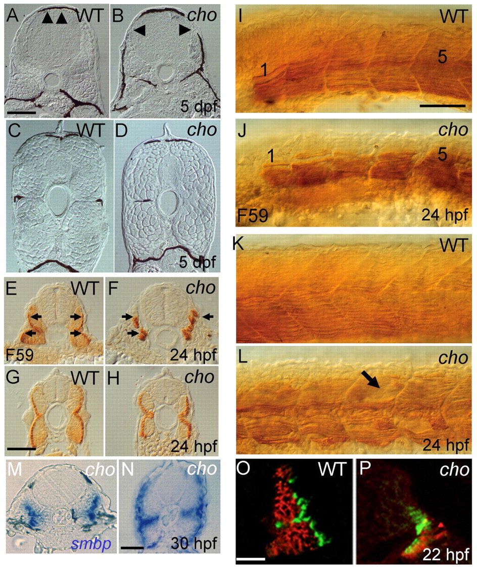
Fig. 4 Somite defects in cho mutants. (A-D) Transverse sections of 5 dpf embryos reveal defective muscle block extension in anterior (arrowheads, A,B), but not posterior (C,D), trunk of cho mutants (B,D) as compared with WT (A,C). cho mutants typically lack melanophores and horizontal myoseptum; exceptionally, as here (left side), a single melanophore may be present, but in an abnormal position. (E-H) Sections of anterior (E,F) and posterior (G,H) trunk of 24 hpf WT (E,G) and cho mutant (F,H) embryos labelled with F59 antibody to show slow muscle fibres (arrows in E,F). (I-L) Lateral view of anterior (I,J; somites 1 and 5 indicated) and posterior (K,L) trunk of 24 hpf WT (I,K) and mutant (J,L) embryos labelled with F59 antibody; note the disorganised slow muscle fibres and apparent holes in fibre pattern (arrow) in cho mutant. (M,N) Transverse sections of anterior (M) and posterior (N) trunk of 30 hpf cho mutant showing smbp RNA in situ hybridisation. (O,P) At the 26-somite stage, WT somites (O) consist largely of fast muscle (red; antibody EB165) under surface slow muscle (green; BAD5), whereas cho mutants (P) displayed severely decreased fast muscle. Scale bars: A-D,I-L, 75 μm; E-H, 25 μm; M,N, 40 μm; O,P, 25 μm.