
|
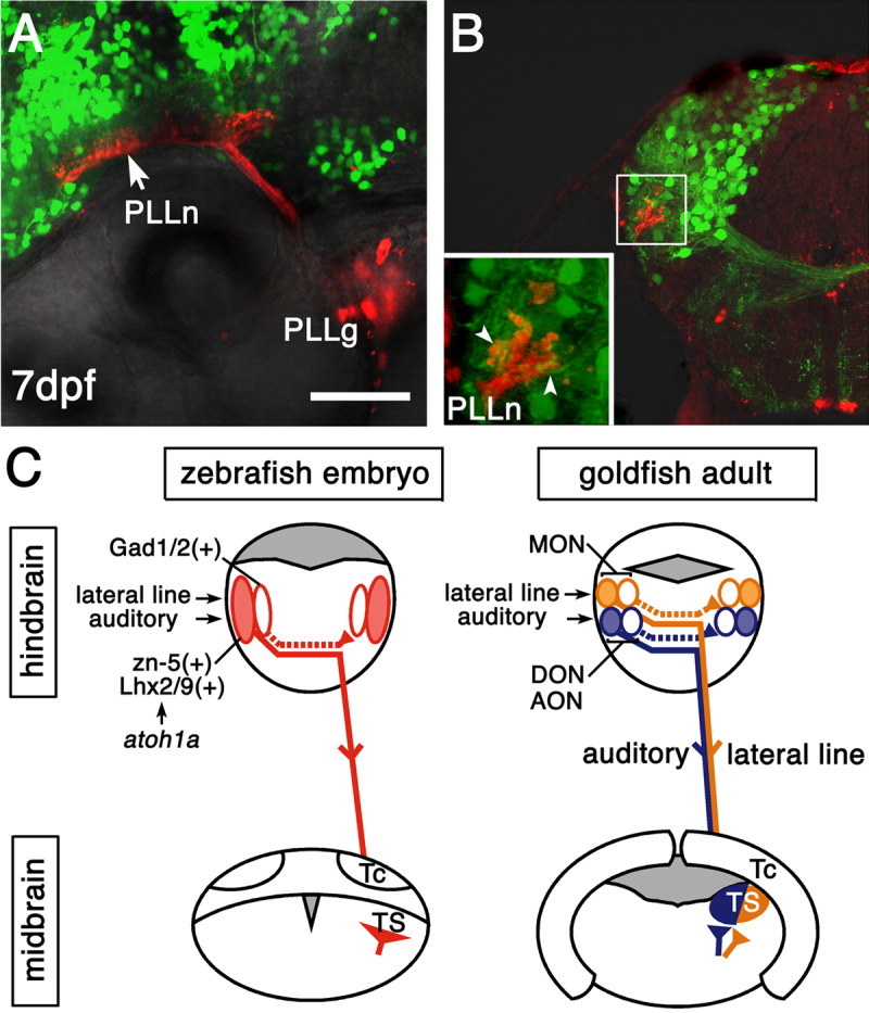
Fig. 5 The zn-5(+)Lhx2/9(+) and Gad1/2(+) neurons might receive auditory and lateral line information. A,B: Posterior lateral line nerves (PLLn) were labeled by tetramethylrhodamine-dextran in a 7 days postfertilization (dpf)Tg(zic1:Gal4VP16/UAS:GFP) embryo. PLLg, posterior lateral line ganglia. A: Lateral view, dorsal is to the top, anterior is to the left. The central nerves of the posterior lateral line enter the hindbrain, then bifurcate and extend longitudinally in the dorsal hindbrain (Alexandre and Ghysen,[1999]), where the green fluorescent protein-positive (GFP(+)) neurons are abundant. B: Coronal section, dorsal is to the top, lateral is to the left. Most of the GFP(+) neurons are located dorsomedially to the axon terminals of the PLLn. Many GFP(+) neuropiles extending toward the terminals of the PLLn were observed (inset, arrowheads). This organization at the larval stage is similar to that of the adult zebrafish and goldfish (New et al.,[1996]; Wullimann et al.,[1996]). C: Schematic illustration of the connectivity between the octaval and octavolateral nuclei and torus semicircularis (TS) in zebrafish embryos (left) and in adult goldfish (right). In the dorsolateral hindbrain of zebrafish embryo (left), at least two distinct classes of neurons are distinguished by the cell locations, molecular profiles, and projection patterns. The commissural axons of the zn-5(+)Lhx2/9(+) neurons (red, filled ovals) form the lateral longitudinal fascicle and project to the midbrain TS. They likely contribute to both the auditory-related and lateral line-related octaval and octavolateral nuclei. It remains to be determined, however, whether they are already segregated in embryos or segregated later at an adult stage (compare with the illustration of adult goldfish on the right). The commissural axons of the Gad1/2(+) neurons (red, open ovals) project to the contralateral hindbrain. For simplicity, projections only from the left side are shown. The bHLH factor Atoh1a is specifically required for development of the zn-5(+)Lhx2/9(+) neurons but not for development of the Gad1/2(+) neurons. In adult goldfish (right), the ascending projection neurons (yellow, filled oval and solid line) in the medial octavolateral nucleus (MON) provide lateral line information to the lateral region of the midbrain TS (yellow; McCormick and Hernandez,[1996]; New et al.,[1996]). There is also a projection to the contralateral MON (yellow, open oval and dotted line; New et al.,[1996]). It is unknown whether distinct neuronal populations project separately to the TS or contralateral MON, or the same neuronal population projects to both the TS and contralateral MON (New et al.,[1996]). The ascending projection neurons (blue, filled oval and solid line) in the descending octaval nucleus (DON) provide auditory information to the medial region of the TS (blue) in goldfish (McCormick and Hernandez,[1996]; Yamamoto and Ito,[2005]). Similarly to the MON, there is also a projection to the contralateral DON (blue, open oval and dotted line; Yamamoto and Ito,[2005]). In other fish species such as carp and catfish, the anterior octaval nucleus (AON) also provides auditory information to the medial region of the TS (Echteler,[1984]; Yamamoto and Ito,[2005]). In goldfish, this nucleus seems to provide few, if any, projections to the TS (McCormick and Hernandez,[1996]; Yamamoto and Ito,[2005]). UAS, upstream activating sequences. Scale bar = 50 μm.