
IMAGE
Fig. 1
- ID
- ZDB-IMAGE-070309-1
- Genes
- Publication
- Wilson et al., 2007 - Cadherin-4 plays a role in the development of zebrafish cranial ganglia and lateral line system
- All Figures
- Figures for Wilson et al., 2007
Image
|
Figure Caption
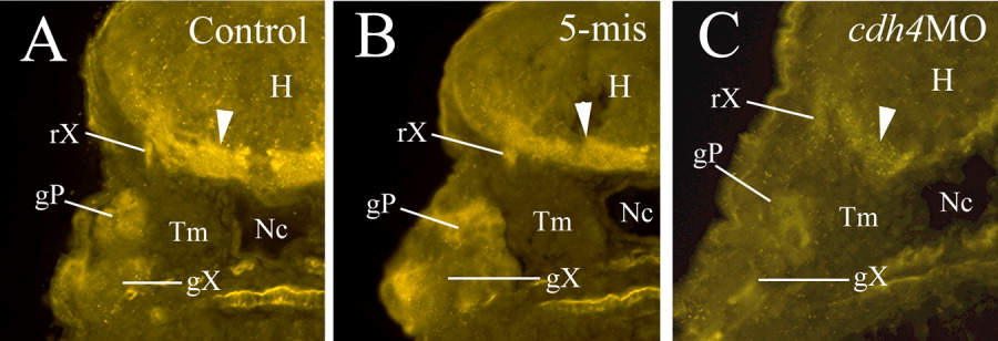
Fig. 1 Cdh4 immunostaining showing that Cdh 4 protein expression levels are greatly reduced in a cdh4 morphant injected with RcadMphA (cdh4MO, C) compared to a control embryo (A) or a 5-mis MO-injected embryo (5-mis, B). All panels are cross-sections in the hindbrain region (dorsal is up) at the level of posterior lateral line ganglion and vagal ganglion (55 hpf). The arrowhead points to Cdh4 immunoreactive fiber tracts in the ventral hindbrain. gP, posterior lateral line ganglion; gX, vagal ganglion; H, hindbrain; Nc, notochord; rX, vagal root; Tm, trunk muscles.
Figure Data
Acknowledgments
This image is the copyrighted work of the attributed author or publisher, and
ZFIN has permission only to display this image to its users.
Additional permissions should be obtained from the applicable author or publisher of the image.
Full text @ Dev. Dyn.