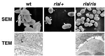
Fig. 6 Cell membrane and microtubule marginal band defects of ris erythrocytes. SEM and TEM analysis of peripheral blood cells. Wild-type erythrocytes are elliptical with smooth biconcave membrane surface. A central nuclear bulge (arrow) located within the biconcavity of wildtype erythrocytes corresponds to the nucleus. Heterozygote ris/+ erythrocytes are elliptical, but the membrane surface appears irregular (asterisk) and biconcavity is lost (arrowheads). Homozygote ris/ris red cells are smaller, spherocytic, and show profound surface pitting and spiculated membrane projections (asterisk). TEM analysis at 3000x magnicication, shows that the MB is situated adjacent to the lipid bilayer (arrows). Wild-type or ris/+ (not shown) MB consists of 20-24 microtubules, whereas ris MB consists of only 12-14 microtubule filaments.
Image
Figure Caption
Acknowledgments
This image is the copyrighted work of the attributed author or publisher, and
ZFIN has permission only to display this image to its users.
Additional permissions should be obtained from the applicable author or publisher of the image.
Full text @ Development

