
Fig. 7
- ID
- ZDB-IMAGE-061228-6
- Publication
- Zolessi et al., 2006 - Polarization and orientation of retinal ganglion cells in vivo
- All Figures
- Figures for Zolessi et al., 2006
|
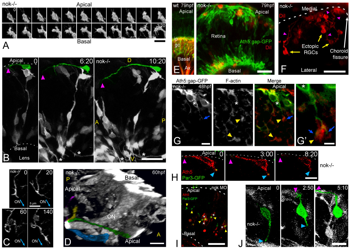
Fig. 7 Retinal ganglion cells (RGCs) polarize, but can be inverted in nok mutants. (a) Complete sequence of images from a three-dimensional (3D) reconstruction, seen from a lateral-apical perspective, of an ectopic RGC in a nok-/- retina. Just before axon outgrowth, the RGC presents several filopodia, mostly extending from the site of axon formation (compare with Figure 2c). Scale bar, 5 μm. (b) Four-dimensional (4D) analysis of a nok-/-embryo injected with ath5:gap-gfp DNA. The ectopic RGC pseudo-colored in green is extending a long neurite (arrowhead) on the retinal outer surface. Time point 0 is at 32 hpf. Asterisks indicate axons from basally located RGCs; A, anterior, D, dorsal; P, posterior; V, ventral. Scale bar, 25 μm. (c) Sequence of 3D reconstructions from a nok-/-embryo expressing ath5:gap-gfp, where an axon growing on the retinal outer surface loops before continuing its growth apparently directed towards the optic nerve (ON) exit. Scale bar, 5 μm. (d) 3D reconstruction taken from a 4D analysis of a nok-/- embryo, transgenic for ath5:gap-gfp. The eye is seen from a medio-ventral position. The pseudo-color highlights four ectopic fascicles of axons joining the optic nerve outside the retina (Additional file 7). Scale bar, 10 μm. (e) Extended-focus confocal images of ath5:gap-gfp transgenic embryos injected with the lipophilic dye DiI into the right tectum to label RGCs retrogradely in the left eye. Scale bar, 15 μm. (f) Ventral view of an eye from a nok-/- embryo in which RGCs have been retrogradely labeled with DiI. The axons of the ectopic RGCs are seen growing towards the optic nerve (not shown in the picture) on the outer retinal surface. Scale bar, 25 μm. (g) Optical section of an ath5:gap-gfp transgenic, nok-/- retina, stained with phalloidin-Texas red. The 'apical' process (blue arrow) of an ectopic ath5:gap-gfp cell (asterisk) ends close to an area where actin filaments appear accumulated (yellow arrowhead). (g' ) Higher magnification of the same cell. Scale bars, 5 μm (g) and 2 μm (g'). (h) Sequence of optical sections from a 4D analysis of a nok-/- retina expressing ath5:gap-rfp and Par3-GFP. The ath5:gap-rfp-positive cell is located close to the apical surface of the retina and is extending a neurite on the outer retinal surface (purple arrowhead). On its other end, another process shows an accumulation of Par3-GFP in its tip (blue arrowhead). Time point 0 is at 34 hpf. Scale bar, 10 μm. (i) Optical section of a retina from a living nok-/- embryo injected with ath5:gap-rfp DNA and par3-gfp mRNA. The Par3-GFP protein is accumulated in apparently random positions inside the retina, and the ath5:gap-rfp-positive cells can be found either on the apical or basal sides of these accumulation points. Scale bar, 20 μm. (j) Sequence of extended-focus images from a 4D analysis of a nok-/-, ath5:gap-gfp transgenic embryo, in which an ectopic differentiating RGC (highlighted in green) is seen to retract a basally directed process and then extend an axon on the retinal outer surface (see Additional file 8 for a rotated 3D reconstruction of this cell). Time point 0 is at 32 hpf. Scale bar, 10 μm. All time stamps are in hours:minutes.