
|
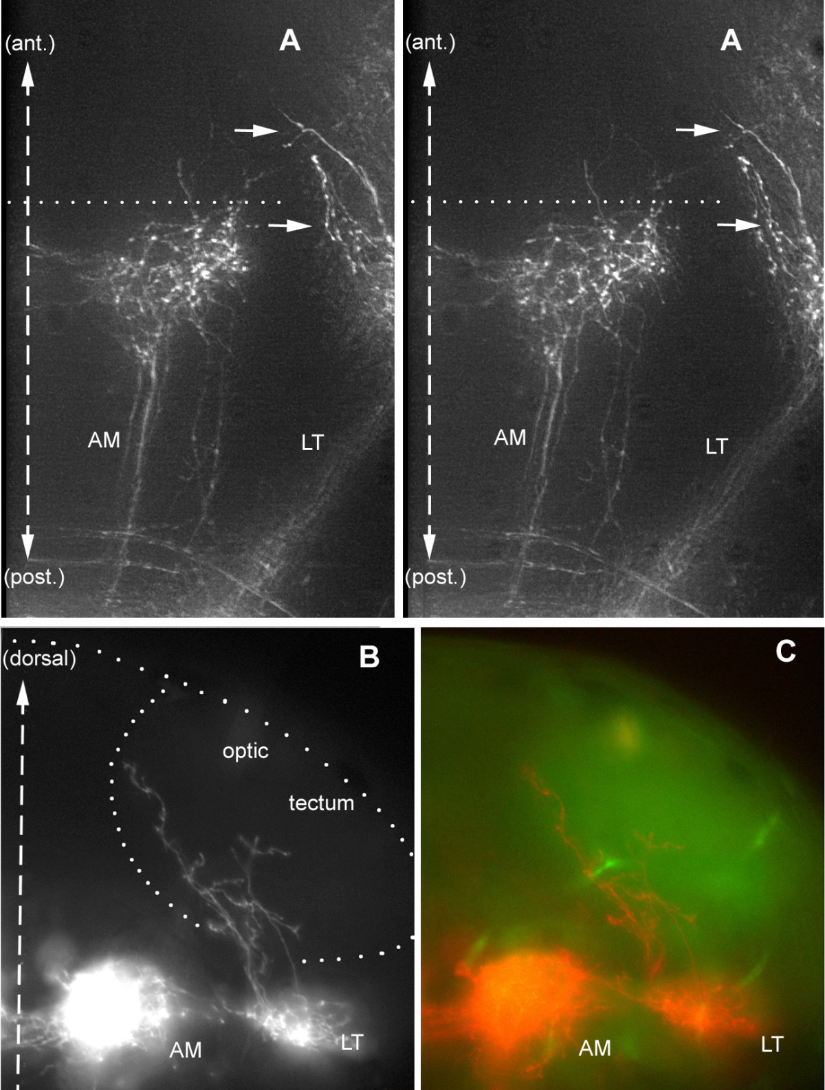
Fig. 7 A few fibers escape from the LT projection and climb into the optic tectum. (a) Dorsal stereo-view of a whole-mount embryo (to be seen with crossed eyes). The lettering has been adjusted to correspond to the dorso-ventral level of the corresponding features; thus, the arrows that point to the climbing fibers will appear much more dorsal to 'AM', whereas 'LT' will appear slightly ventral to 'AM'. (b, c) Climbing fibers seen in a vibratome section. The outline of the tectum in (b) has been drawn based on the autofluorescence pattern seen under blue exciting light (c), which reveals fibrous material (neuropil) as opposed to cell bodies (nuclei).