
Fig. 6
- ID
- ZDB-IMAGE-061206-19
- Publication
- Lee et al., 2006 - Canonical Wnt signaling through Lef1 is required for hypothalamic neurogenesis
- All Figures
- Figures for Lee et al., 2006
|
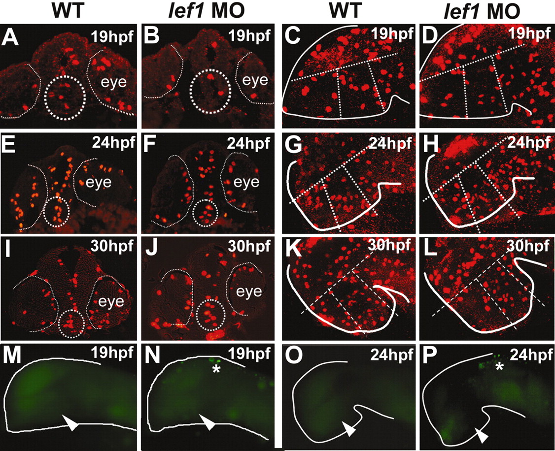
Fig. 6 Analysis of proliferation and apoptosis in lef1 morphants. (A-L) In lef1 morphants, pH3-positive cells are observed in the posterior hypothalamus at 19, 24 and 30 hpf, indicating that proliferating cells are still present in this region. (A,B,E,F,I,J) Cross-sections through posterior hypothalamus (outlined by dotted circles). (C,D,G,H,K,L) Confocal projections of pH3-stained embryos used for quantitative analysis. The central region outlined by broken lines was defined as posterior hypothalamus, based on morphology in bright-field views. The region farthest to the right was excluded from counts because it does not express lef1 and is unaffected in lef1 morphants (Fig. 4Q-T). (M-P) Apoptotic cells identified by TUNEL staining are absent in the posterior hypothalamus at 19 hpf and 24 hpf in wild-type and lef1 MO-injected embryos (arrowheads). Asterisks indicate apoptotic cells in the dorsal brain of lef1 morphants.