
Fig. 9
- ID
- ZDB-IMAGE-061121-21
- Publication
- Eroglu et al., 2006 - Critical role of Brg1 member of the SWI/SNF chromatin remodeling complex during neurogenesis and neural crest induction in zebrafish
- All Figures
- Figures for Eroglu et al., 2006
|
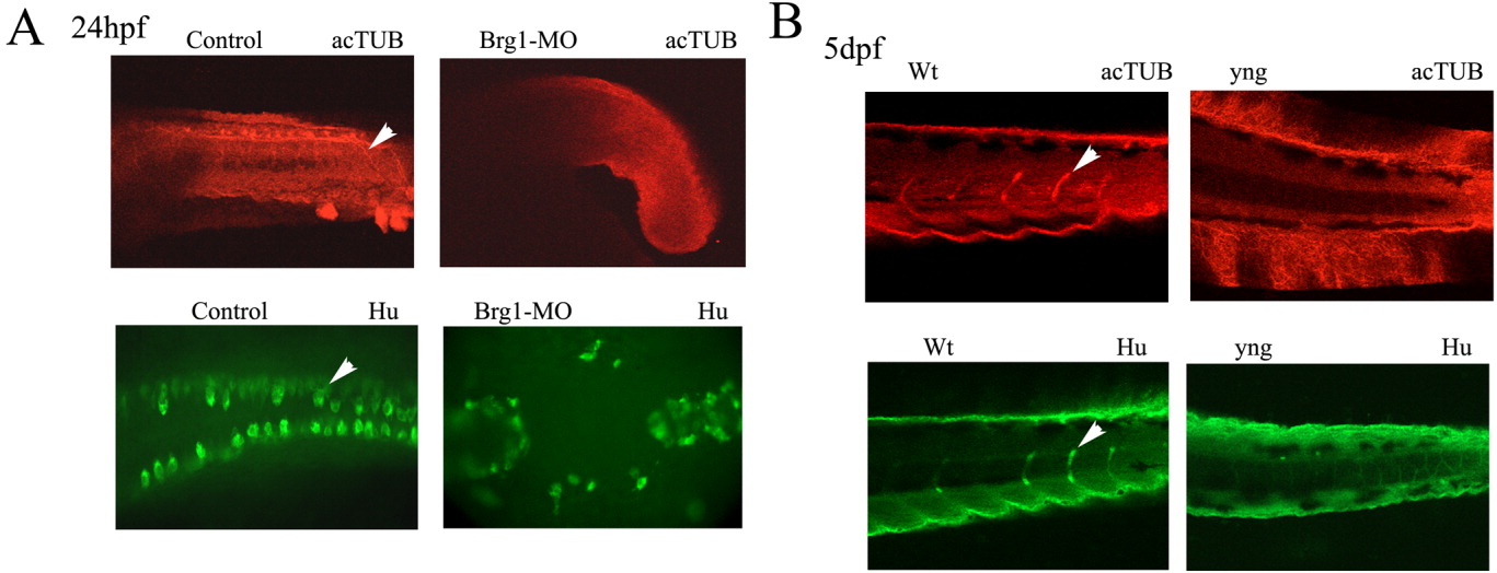
Fig. 9 The Brg1-MO injected and yng mutant embryos exhibit reduced peripheral neurons and axons. A: One- to two-cell stage embryos were injected with control-MO or Brg1-MO, and at 24 hpf embryos were fixed and immunostained with antibodies to acetylated tubulin (acTUB) (lateral) or HuC/D (dorsal). Original magnification 200×. B: The yng mutant embryos were fixed and immunostained at 5 dpf as above. Lateral views are presented. The acTUB and HuC/D were detected using AlexaFluor 594- and AlexaFluor 488- labeled secondary antibodies, respectively. Image analysis was performed with conventional fluorescent or confocal microscopy. In A, note the reduced primary motor nerves and Rohon Beard sensory neurons in Brg1-MO-injected embryos compared to control (arrowheads). In B, note the reduced DRGs in the trunk region of yng mutant embryos compared to wild type indicated by arrowheads. Original magnification 200×.