
Fig. 8
- ID
- ZDB-IMAGE-061121-20
- Publication
- Eroglu et al., 2006 - Critical role of Brg1 member of the SWI/SNF chromatin remodeling complex during neurogenesis and neural crest induction in zebrafish
- All Figures
- Figures for Eroglu et al., 2006
|
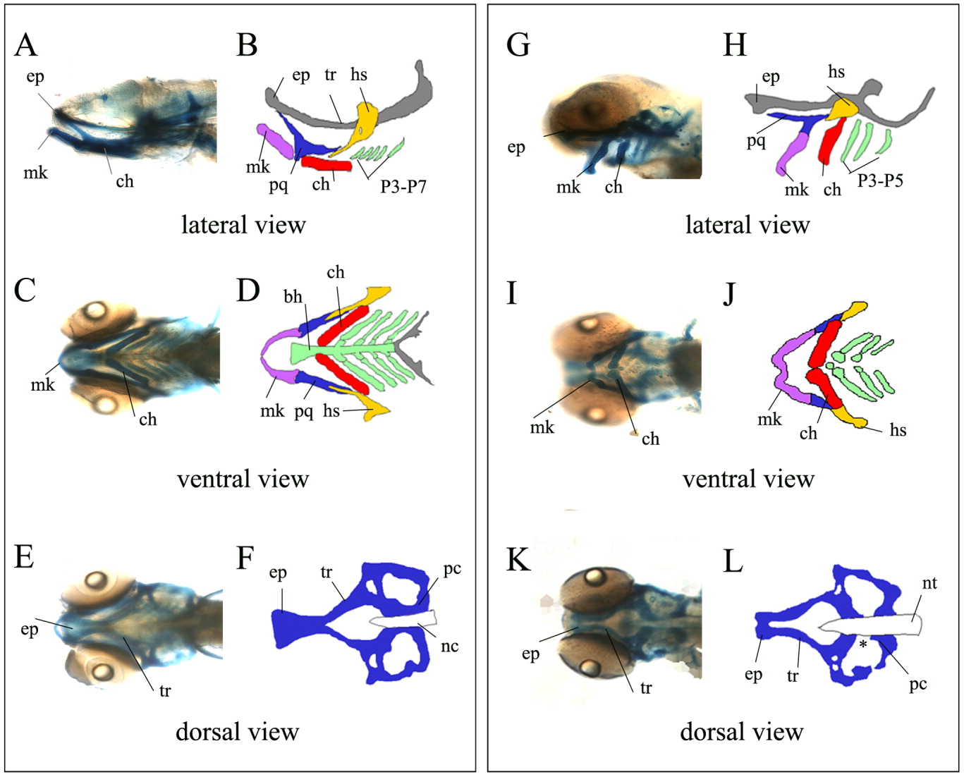
Fig. 8 The yng mutant embryos show defects in neural crest cell differentiation. Craniofacial skeletons and schematic drawings of wild type (A,C,E and B,D,F), and yng mutant (G,I,K and H,J,L), embryos following Alcian Blue staining at 5 dpf. The lateral (A and G) and ventral (C and I) views show that all the pharyngeal cartilages form ventrally and the most posterior arches (P6-P7) are reduced or absent in yng mutant embryos. The dorsal processes of the platoquadrate (pq) fail to articulate with the ethmoid plate (ep). The basihyal (bh) along the midline is reduced. The dorsal view of neurocranium (E and K) shows that neural crest-derived ethmoid plate (ep) and mesodermally derived parachordals (pc) are reduced. bh, basihyal; ch, ceratohyal; ep, ethmoid plate; hs, hyosymplectic; mk, Meckel's cartilage; nc, notochord; pc, parachordal; pq, platoquadrate; tr, trabeculae; Wt, wild-type. Note that eyes were removed from the wild type embryo (A) to observe fine details. Original magnification 200×.