
Fig. 5
- ID
- ZDB-IMAGE-061121-17
- Publication
- Eroglu et al., 2006 - Critical role of Brg1 member of the SWI/SNF chromatin remodeling complex during neurogenesis and neural crest induction in zebrafish
- All Figures
- Figures for Eroglu et al., 2006
|
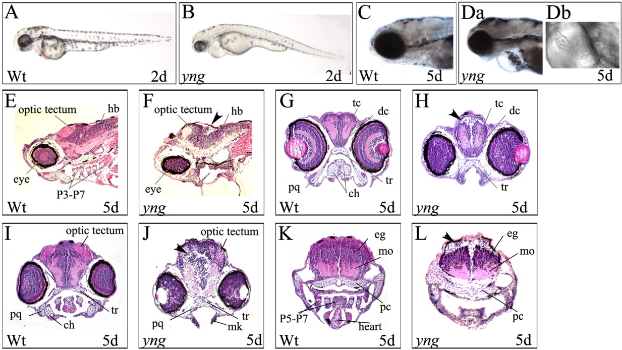
Fig. 5 The yng mutant embryos exhibit multiple defects. A,B: yng mutant embryos exhibit lower expression of melanin compared to wild type at 2 dpf. Original magnification 40×. C,Da,Db: yng mutant embryos exhibit cardiovascular defects compared to wild type at 5 dpf. Original magnification 60×. E-L: yng mutant embryos exhibit neurodevelopmental defects compared to wild type at 5 dpf. E and F are sagittal sections. Arrow indicates position of the cerebellum. G-L are coronal sections showing evidence of cell loss in yng mutant embryo (arrowheads). Histological sections were stained with hematoxylin and eosin. hb, hindbrain; P3-P7, pharyngeal arches 3-7; tc, telencephalon; dc, diencephalons; tr, trabeculae; pq, palatoquadrate; ch, ceratohyal; mk, Meckel's cartilage; P5-P7, pharyngeal arches 5-7; pc, parachordal; mo, medulla oblongata; eg, eminentia granularis. Original magnification 200×.