Image
Figure Caption
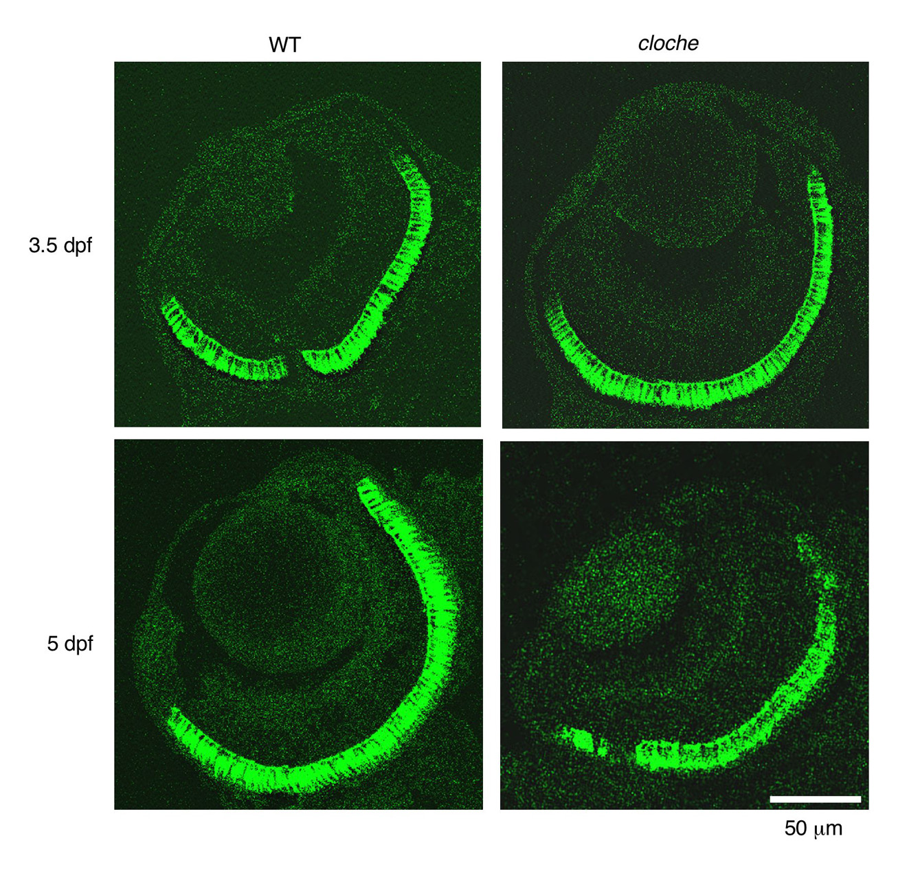
Fig. S1 Retina photoreceptor. Sections were prepared from 3.5 and 5 dpf embryos, and analyzed by immunohistochemistry with antibodies that detect photoreceptors (green). Photoreceptor and retinal thickness are the same in cloche as in wild type. In addition, the photoreceptor structure in cloche does not appear to change from 3.5 to 5 dpf.
Acknowledgments
This image is the copyrighted work of the attributed author or publisher, and
ZFIN has permission only to display this image to its users.
Additional permissions should be obtained from the applicable author or publisher of the image.
Full text @ Development

