
Fig. 7
- ID
- ZDB-IMAGE-060828-13
- Publication
- Raeker et al., 2006 - Obscurin is required for the lateral alignment of striated myofibrils in zebrafish
- All Figures
- Figures for Raeker et al., 2006
|
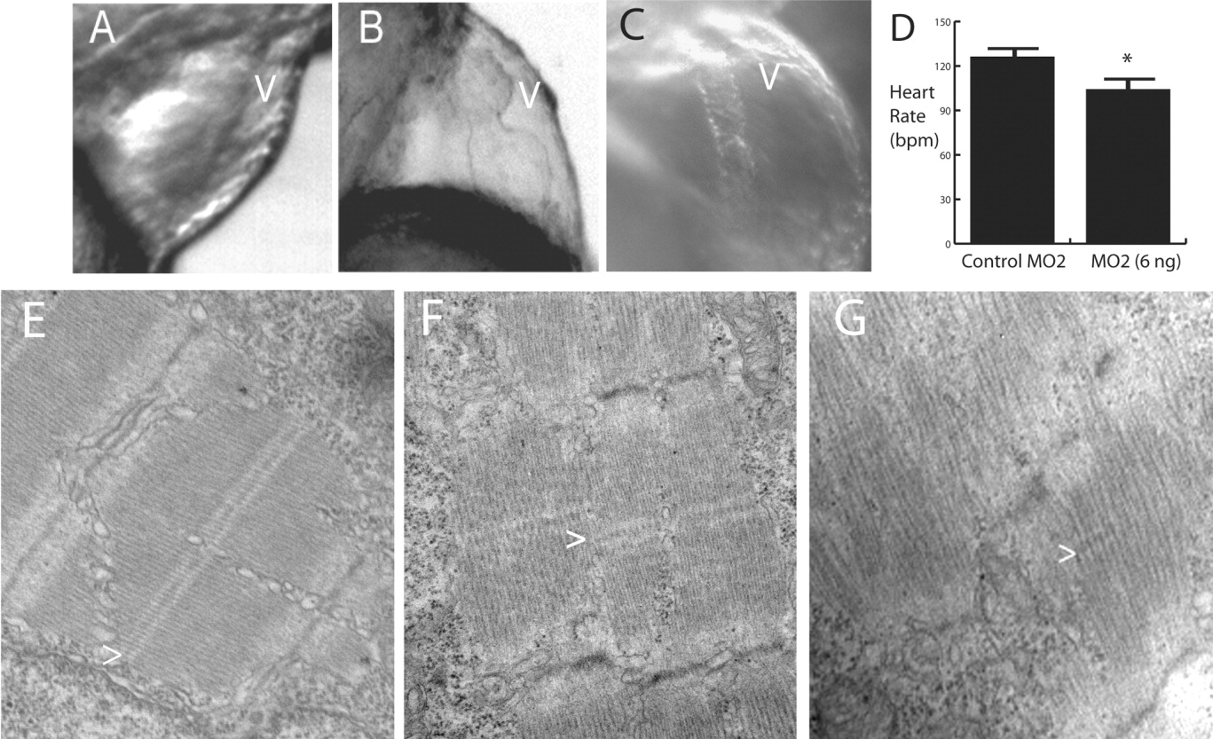
Fig. 7 Cardiac abnormalities in morpholino-treated embryos. A-C: Morphant embryos demonstrated a spectrum of cardiac defects that ranged from mild (B) to severe (C) ventricular (v) hypoplasia. The most severely affected embryos (6 ng of MO2) studied had tube-like hearts (C). A control 72-hpf embryo is shown for comparison (A). All cardiac defects were associated with mild to marked pericardial edema. D: The heart rate of the morphant embryos (6 ng of MO2) was significantly less than that of the control embryos (*t-test; P < 0.01). As with skeletal muscle, cardiac myofibrils in morphant embryos [3 ng (F) and 6 ng (G) of MO2] were poorly aligned and organized compared to those in control-injected embryos (E). Some M bands (>) are apparent in morphant embryos but are much more irregular than in control embryos.