
Image
|
Figure Caption
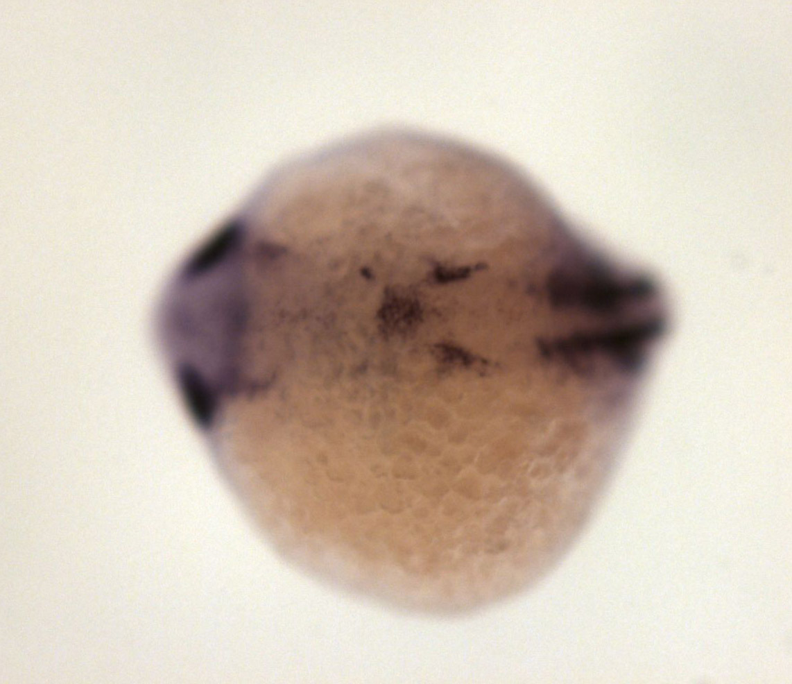
Fig. 3 ventral diencephalon, posterior part of retina, epiphysis, cranial ganglia, dorsal hindbrain, ventral and dorsal somites
Orientation
| Preparation | Image Form | View | Direction |
| whole-mount | still | dorsal | anterior to left |
Figure Data