
|
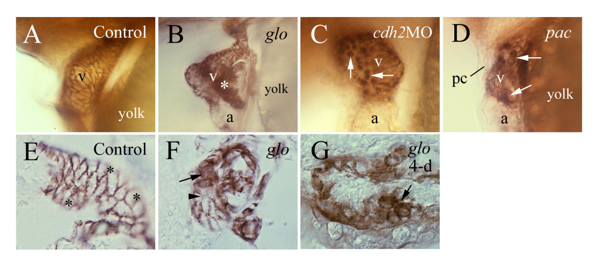
Fig. 6 Zn-5 immunostaining reveals myocardiocyte defectsin a cadherin2 morphant, a pac mutant embryo and glo mutant embryos. Zn-5 immunostaining reveals myocardiocyte defects in a cadherin2 morphant (panel C), a pac mutant embryo (panel D) and glo mutant embryos (panels B, F and G). All embryos, except panel G (4-day old) are 48–50 hpf. Panels A-D are lateral views of whole mount hearts with anterior to the left and dorsal up. Panels E-G are sections of zebrafish hearts processed for whole mount Zn-5 immunostaining. The asterisk in panel B indicates a large cluster of myocardiocytes with Zn-5 immunoreactivity detected in both their cell membranes and cytoplasm. Arrows in panels C and D point to round shaped myocardiocytes with staining in the cell membrane and cytoplasm. Panel E is a parasagittal section (anterior to the left lower corner and dorsal to the left upper corner) of a control heart. Asterisks in this panel indicate regions that are out of focus. Panel F is a parasagittal section of a glo mutant heart (anterior to the left and dorsal up). Panel G is a parasagittal section (anterior to the left and dorsal up) of 4-day old glo mutant heart. Arrows in panels F and G point to round shaped myocardiocytes with labeling in their cell membranes and cytoplasm, while the arrowhead in panel F indicates a myocardial cell with weak labeling on its cell membrane.