
|
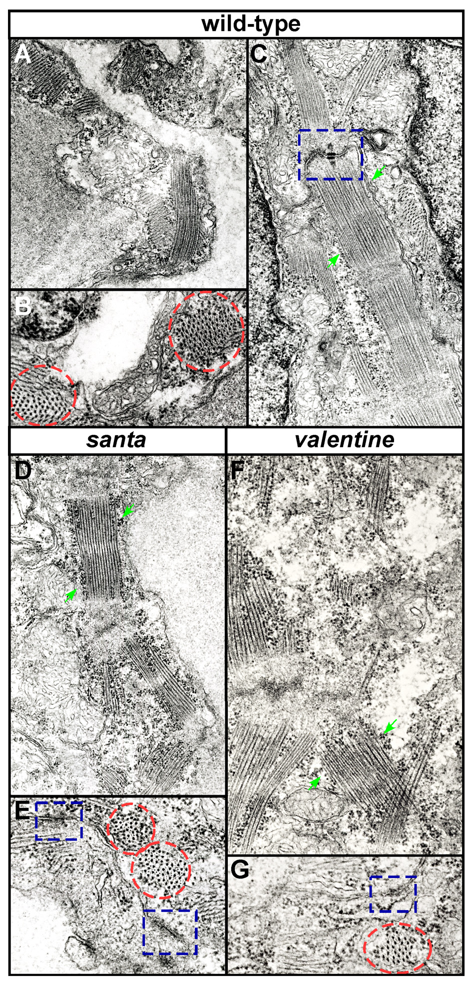
Fig. S2 Sarcomeres are present in the hearts of san and vtn mutant embryos. Transmission EM shows sarcomeres (green arrows) in the hearts of wild-type (A-C), san (D,E) and vtn (F,G) embryos, but with some disarray in the mutants (D,F). Sarcomeres are shown in cross-section (indcated by red circles) in wild-type (B), san (E) and vtn (G) mutants. Both san and vtn mutant hearts are still able to contract although circulation is not generated suggesting that actin/myosin arrangement is normal. Intercalated discs (indicated by blue boxes) are detectable in both mutants (E,G) although their structure in wild-type hearts is better defined (C). Comparison with similar EM analyses of cardiac tissue suggests the electron-dense material along and between the myofilaments is glycogen.