
|
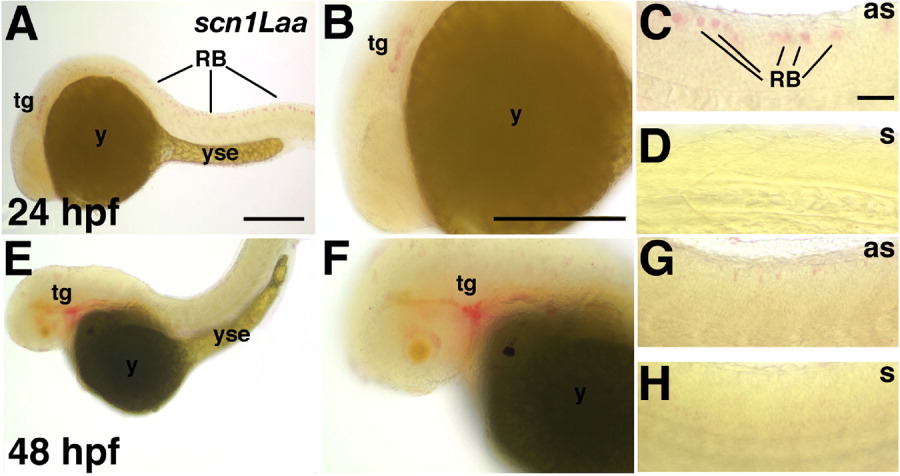
Fig. 2 Scn1Laa is expressed in sensory neurons of the peripheral nervous system and Rohon-Beard (RB) cells. A-D: At 24 hours postfertilization (hpf), scn1Laa mRNA is detected in the trigeminal ganglion and RB cells. A: A low-magnification view reveals expression in an anterior domain caudal to the eye and in RB cells throughout the spinal cord. B: At higher magnification, the anterior expression domain is recognized as the developing trigeminal ganglion migrating rostrally toward the eye. C: The posterior expression is found in RB cells of the spinal cord. D: The sense probe (s) does not reveal a signal under the same conditions used for the antisense probe (as). E-H: At 48 hpf, in situ hybridization signals are stronger in the trigeminal ganglion but weaker posteriorly in the spinal cord. E,F: At 48 hpf, the trigeminal ganglia have migrated to their characteristic position just caudal to the eye and continue to express scn1Laa. G: At 48 hpf, dorsal RB cells continue to express scn1Laa transcripts. H: The sense probe reveals no hybridization signal. as, antisense probe; n, notochord; RB, Rohon-Beard cell; s, sense probe; tg, trigeminal ganglion; yse, yolk sac extension. In this and subsequent figures, whole-mount photos are oriented with anterior to the left and dorsal up. Scale bars = 250 μm in A (applies to A,E), 100 μm in B (applies to B,F), 100 μm in C (applies to C,D,G,H).