
Fig. 4
- ID
- ZDB-IMAGE-060707-4
- Publication
- Goishi et al., 2006 - {alpha}A-crystallin expression prevents {gamma}-crystallin insolubility and cataract formation in the zebrafish cloche mutant lens
- All Figures
- Figures for Goishi et al., 2006
|
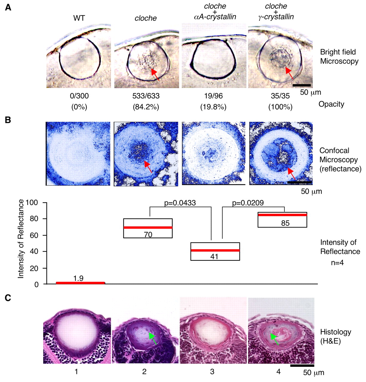
Fig. 4 αA-crystallin expression prevents cataract formation in cloche. αA- (panel 3) or γ-crystallin (panel 4) mRNA was injected into embryos at the one- to four-cell stage and phenotypes were compared with uninjected wild type (panel 1) and cloche lens (panel 2). (A) Living embryos were analyzed by stereomicroscopy and the number of opaque lenses was counted. Red arrows point to scattered light. The percent of lenses that demonstrate opacity is shown in parentheses. (B) Living embryos lenses were analyzed by confocal microscopy and the intensity of reflected light was measured. Red arrows indicate areas of reflected light. The median reflectance was measured as in Fig. 3D. The Mann-Whitney U test was used to test for differences between two groups. The differences in the lens between cloche embryos and cloche embryos overexpressing αA-crystallin (P=0.0433) and the differences between cloche overexpressing αA-crystallin versus γ-crystallin (P=0.0209) are significant. (C) Sections were prepared from 4 dpf embryos and analyzed by Hematoxylin and Eosin staining. The green arrows indicate nuclei. The number of lens fiber cell nuclei per section was analyzed at 4 dpf. Three sections of each lens were analyzed by an image analysis program (ImageJ). Fourteen embryos were analyzed. At 4 dpf, wild type and cloche had 0.0±0.0 and 22.6±5.0 nuclei, respectively. In the rescue experiment, αA-crystallin overexpression reduced the nuclei from 22.6±5.0 to 3.0±0.0 (mean±s.d.).