
|
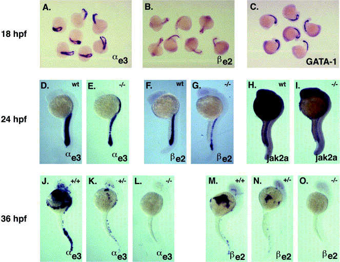
Fig. 5 Analysis of globin gene expression in zin mutants by whole-mount in situ hybridization analysis. At 18 hpf, no defect in the expression of αe3-globin (A), βe2-globin (B), or GATA-1 (C) can be detected in clutches arising from an intercross of two zin AB/SJD heterozygotes. GATA-1 is shown as a control for erythroblast number; at this stage, no cell lysis is apparent in zin mutant embryos. At 24 hpf, expression of αe3-globin (D, E) and βe2-globin (F, G) is reduced in zin homozygotes. This effect is likely to be due to a decrease in red blood cell number, as shown by reduced expression of the erythroid marker jak2a in zin homozygotes (H, I). By 36 hpf, levels of αe3-globin and βe2-globin mRNA can be used to distinguish wild-type siblings (J, M), zin heterozygotes (K, N), and zin homozygotes (L, O). The reduction in staining in the mutant embryos is reflective of the severity of the anemia at this stage and represents a secondary effect of cell lysis.
Reprinted from Developmental Biology, 255(1), Brownlie, A., Hersey, C., Oates, A.C., Paw, B.H., Falick, A.M., Witkowska, H.E., Flint,J., Higgs, D., Jessen, J., Bahary, N., Zhu, H., Lin, S. and Zon, L., Characterization of embryonic globin genes of the zebrafish, 48-61, Copyright (2003) with permission from Elsevier. Full text @ Dev. Biol.