Image
Figure Caption
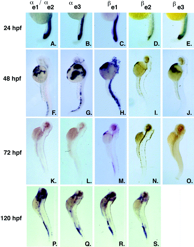
Fig. 3 Whole-mount in situ hybridization analysis of embryonic globin gene expression in the zebrafish. Expression of the embryonic globin genes αe1/αe2 (A, F, K, P), αe3 (B, G, L, Q), βe1 (C, H, M, R), βe2 (D, I, N, S), and βe3 (E, J, O) were examined at 24 (A–E), 48 (F–J), 72 (K–O), and 120 hpf (P–S) by whole-mount in situ hybridization. The sequence of the αe1- and αe2-globins is highly similar, and so it is unlikely that these mRNAs can be distinguished by using this technique. The expression of βe3-globin has not been examined at 120 hpf.
Acknowledgments
This image is the copyrighted work of the attributed author or publisher, and
ZFIN has permission only to display this image to its users.
Additional permissions should be obtained from the applicable author or publisher of the image.
Reprinted from Developmental Biology, 255(1), Brownlie, A., Hersey, C., Oates, A.C., Paw, B.H., Falick, A.M., Witkowska, H.E., Flint,J., Higgs, D., Jessen, J., Bahary, N., Zhu, H., Lin, S. and Zon, L., Characterization of embryonic globin genes of the zebrafish, 48-61, Copyright (2003) with permission from Elsevier. Full text @ Dev. Biol.

