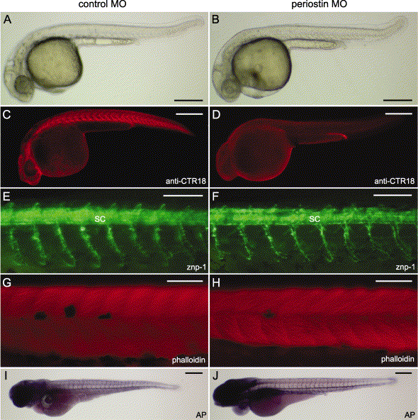
Fig. 5 Characterization of the periostin morphant phenotype. Control MO (A, C, E, G, and I) and periostin MO (B, D, F, H, and J) -injected embryos are shown with the anterior aspect at the left and the dorsal one at the top. (A an B) Live images show morphological phenotypes at 33 hpf. (C and D) Whole-mount immunostaining with the anti-periostin antibody (anti-CTR18) indicates the loss of periostin at the transverse myosepta in the 27-hpf periostin morphants (D). (E and F) Whole-mount immunostaining with znp-1 reveals the primary motor axons of 30-hpf embryos. (G and H) Alexa Fluor 568-phalloidin staining of 48-hpf embryos shows somitic muscles of the midtrunk region. (I and J) Endogenous alkaline phosphatase activities reveal metameric patterns of the intersomitic vessels in 72-hpf embryos. Abbreviations: AP, alkaline phosphatase; MO, morpholino antisense oligonucleotide; SC, spinal cord. Scale bars: A–D, I, and J, 250 μm; E–H, 100 μm.
Reprinted from Developmental Biology, 267(2), Kudo, H., Amizuka, N., Araki, K., Inohaya, K., and Kudo A., Zebrafish periostin is required for the adhesion of muscle fiber bundles to the myoseptum and for the differentiation of muscle fibers, 473-487, Copyright (2004) with permission from Elsevier. Full text @ Dev. Biol.

