
|
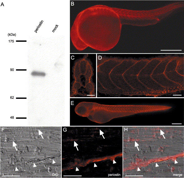
Fig. 3 Localization of periostin protein in embryos and adult muscles. (A) Western blot analysis of the conditioned medium from supernatants of periostin transfectants or mock transfectants by using antiperiostin antibody (anti-CTR18). An immunoreactive band of 86 kDa is observed in the lane of periostin transfectants, which size corresponds to the calculated molecular weight of 85,884. (B–F) Whole-mount immunostaining with the anti-periostin antibody. Periostin staining of 24-hpf embryos (B–D) in transverse (C) and sagittal (D) sections of the trunk reveals the localization of periostin in the transverse myosepta and the periphery of myotomes, respectively. In 48-hpf embryos (E), periostin localization in the transverse myosepta is maintained. (F, G, and H) Immunohistochemistry of skeletal muscle of 9-month-old zebrafish with cryosectioning. DIC microscopic image reveals that striated muscle fibers and a myoseptum (white arrowheads) diagonally attach each other (F). Periostin immunoreactivities are present in the myoseptum (white arrowheads) and adjoining sites among the muscle bundles (G, H, and white arrows). (H) Merged image of DIC and fluorescent images. Abbreviation; DIC, differential interference contrast microscope. Scale bars: B and E, 250 μm; C, D, and F–H, 25 μm.
Reprinted from Developmental Biology, 267(2), Kudo, H., Amizuka, N., Araki, K., Inohaya, K., and Kudo A., Zebrafish periostin is required for the adhesion of muscle fiber bundles to the myoseptum and for the differentiation of muscle fibers, 473-487, Copyright (2004) with permission from Elsevier. Full text @ Dev. Biol.