Image
Figure Caption
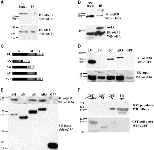
Fig. 4 Mib is associated with Delta in Cos7 cells. (A, B) Western blot analysis of co-immunoprecipitation samples reveals the association between full-length Mib with DeltaD (upper panel) and HA-DeltaAICD (lower panel). (C) Schematic diagram of the Mib portion of EGFP fusion proteins. The solid vertical bars indicate the ankyrin repeats, the empty triangles indicate the ring fingers. (D, E) Co-IP analysis of Mib deletions and DeltaD shows that the N-terminal half is necessary for DeltaD association. (F) GST pull-down analysis shows that the N-terminal half is sufficient to associate with DeltaD.
Acknowledgments
This image is the copyrighted work of the attributed author or publisher, and
ZFIN has permission only to display this image to its users.
Additional permissions should be obtained from the applicable author or publisher of the image.
Reprinted from Developmental Biology, 267(2), Chen, W., and Casey Corliss, D., Three modules of zebrafish Mind bomb work cooperatively to promote Delta ubiquitination and endocytosis, 361-373, Copyright (2004) with permission from Elsevier. Full text @ Dev. Biol.

