
|
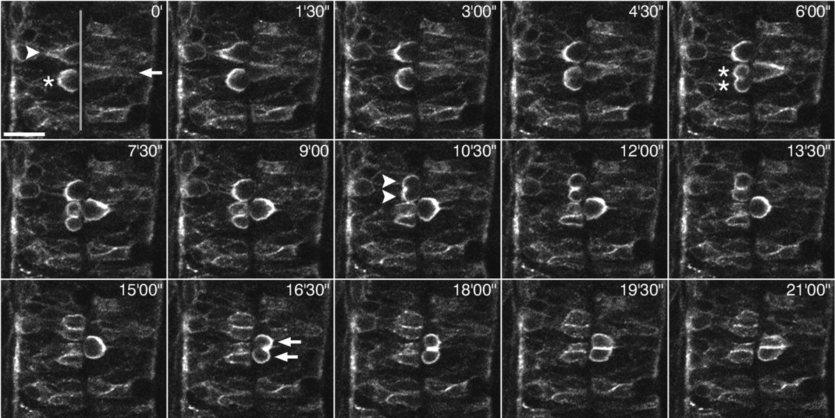
Fig. 5 A sequence of 15 confocal micrographs of a dorsal view of the neural tube of a ~24-hpf-old embryo injected with numb(PTBL PRRL):egfp mRNA. The transparent grey line in the first picture marks the neurocoel. Scale bar = 20 μm. Three cells (arrows, arrowheads, and asterisks in 0') one after another round up at the neurocoel and subsequently undergo mitotic cell division. Note that Numb:EGFP becomes localized to the basolateral cell cortex in all three cells, whereas the apical side is completely devoid of signal. Although Numb:EGFP is clearly asymmetrically localized in these cells, it is distributed to both daughter cells upon division, since the cells divide parallel to the plane of the neuroepithelium. See also Supplemental Material for the corresponding Supplementary Movie 4.